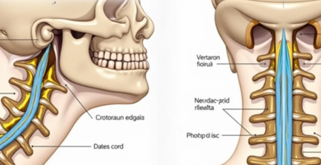
The cervical spine represents one of the most intricate and vital regions of the human skeletal system, with the C3–C4 level serving as a critical junction point for neurological function and structural stability. This mid-cervical segment plays an indispensable role in supporting head movement, protecting essential nerve pathways, and maintaining the delicate balance between mobility and stability that defines optimal spinal health. Understanding the complexities of this anatomical region becomes increasingly important as cervical spine disorders continue to affect millions of individuals worldwide, with degenerative changes at the C3–C4 level representing a significant proportion of cervical pathology cases.
Anatomical structure and vertebral components of C3–C4 cervical segments
The C3 and C4 vertebrae exhibit the characteristic features of typical cervical vertebrae, distinguished by their unique anatomical adaptations that facilitate both structural support and extensive range of motion. These vertebrae possess smaller vertebral bodies compared to their thoracic and lumbar counterparts, reflecting the reduced weight-bearing demands of the cervical region whilst accommodating the complex neurological structures that traverse this area.
Atlas-axis complex relationship to C3–C4 functional units
The relationship between the upper cervical complex (C1–C2) and the C3–C4 segments creates a sophisticated biomechanical system that enables the remarkable mobility of the human neck. Unlike the specialised atlas and axis vertebrae above, the C3–C4 segments begin the transition toward more conventional vertebral architecture whilst maintaining the cervical spine’s characteristic flexibility. This transition zone experiences unique stress patterns that influence both normal function and pathological processes.
The positioning of C3–C4 below the atlantooccipital and atlantoaxial joints means these segments must compensate for forces transmitted from the highly mobile upper cervical region. This compensation mechanism involves complex interactions between the vertebral bodies, facet joints, and supporting ligamentous structures, creating a biomechanical environment that is both resilient and vulnerable to degenerative changes over time.
Cervical lordosis biomechanics at third and fourth vertebrae
The natural cervical lordosis reaches a critical inflection point at the C3–C4 level, where the curvature begins its gradual transition toward the more pronounced lordotic curve of the lower cervical spine. This curvature serves multiple biomechanical functions, including load distribution, shock absorption, and maintenance of optimal spinal alignment for neurological protection. The precise angulation at this level influences stress distribution patterns throughout the entire cervical column.
Alterations in cervical lordosis at the C3–C4 level can significantly impact the biomechanical function of adjacent segments, potentially accelerating degenerative processes or creating compensatory stress patterns that affect overall cervical spine health. Modern lifestyle factors, particularly prolonged forward head postures associated with computer work and mobile device usage, place considerable stress on the natural lordotic curve at this level.
Intervertebral disc morphology between C3–C4 segments
The intervertebral disc at the C3–C4 level exhibits distinct morphological characteristics that reflect the unique functional demands of this spinal segment. The disc consists of a fibrous outer layer (annulus fibrosus) surrounding a gel-like inner core (nucleus pulposus), with the cervical discs being proportionally thinner than those in the lumbar spine whilst maintaining remarkable flexibility.
The C3–C4 disc experiences complex loading patterns during neck movement, with flexion, extension, lateral bending, and rotational forces creating multidirectional stress patterns within the disc structure. These loading characteristics contribute to specific patterns of disc degeneration that are commonly observed at this level, including posterior disc bulging and annular fissuring that can compromise neural structures.
Facet joint orientation and zygapophyseal architecture
The facet joints at the C3–C4 level exhibit a characteristic orientation that facilitates the extensive range of motion required for normal cervical function. These synovial joints are oriented approximately 45 degrees from the transverse plane, allowing for smooth gliding movements during flexion, extension, and coupled lateral bending with rotation. The articular surfaces are covered with hyaline cartilage and surrounded by a joint capsule that provides both stability and nutritional support.
The zygapophyseal joints at this level are particularly susceptible to degenerative changes due to their high mobility and the forces they must withstand during daily activities. Facet joint arthritis at the C3–C4 level can contribute to neck pain, stiffness, and referred pain patterns that may extend into the shoulder and arm regions.
Transverse foramen vertebral artery pathway configuration
The transverse foramina of the C3 and C4 vertebrae provide passage for the vertebral arteries, which play a crucial role in posterior circulation to the brainstem and cerebellum. The configuration of these foramina at the C3–C4 level creates potential sites of vascular compromise in the presence of degenerative changes, osteophyte formation, or traumatic injury.
Understanding the relationship between spinal pathology and vertebral artery function at this level becomes particularly important when considering surgical interventions or evaluating symptoms that may have a vascular component. The anatomical variability in vertebral artery course and diameter at the C3–C4 level requires careful consideration during diagnostic imaging and treatment planning.
Neurological pathways and spinal cord vulnerability at C3–C4 level
The neurological significance of the C3–C4 level cannot be overstated, as this region houses critical neural pathways that control essential bodily functions, including respiratory function, upper extremity motor control, and sensory processing. The spinal cord at this level maintains its cylindrical configuration whilst beginning the gradual expansion that characterises the cervical enlargement, making it particularly vulnerable to compressive forces.
Cervical spinal cord diameter reduction patterns
The spinal cord diameter at the C3–C4 level represents a transitional zone where the cord begins to expand in preparation for the cervical enlargement that extends from C5 to T1. This anatomical configuration creates a relative narrowing that can become clinically significant in the presence of canal stenosis or compressive pathology. The cross-sectional area of the spinal cord at this level typically measures between 60-70 square millimetres, providing limited space for accommodation of compressive forces.
Research indicates that even minor reductions in canal diameter at the C3–C4 level can result in significant neurological symptoms due to the critical nature of the neural pathways traversing this region. The concept of functional reserve becomes particularly important at this level, where the margin for error between normal function and neurological compromise is relatively narrow.
C3–C4 nerve root dermatomal distribution mapping
The nerve roots emerging at the C3–C4 level provide innervation to specific dermatomal and myotomal distributions that have important clinical implications for diagnosis and treatment. The C3 nerve root primarily innervates the posterior and lateral neck regions, with some overlap into the occipital area, whilst the C4 nerve root provides sensation to the shoulder region and contributes to motor innervation of the diaphragm through the phrenic nerve.
Understanding the precise dermatomal patterns associated with C3–C4 nerve root function enables healthcare providers to accurately localise pathology and develop targeted treatment strategies. Nerve root compression at this level can produce characteristic pain patterns that may be confused with other cervical or shoulder pathologies without proper neurological assessment.
Phrenic nerve origin from C3–C4–C5 nerve roots
The phrenic nerve, which provides motor innervation to the diaphragm, receives contributions from the C3, C4, and C5 nerve roots, with the C4 component typically representing the largest contribution. This anatomical relationship makes the C3–C4 level critically important for respiratory function, as pathology at this level can potentially compromise breathing capacity.
The phrenic nerve’s dependence on cervical nerve root function at the C3–C4 level underscores the life-threatening potential of high cervical spinal cord injuries and the importance of maintaining neural integrity at this level.
Clinical assessment of phrenic nerve function becomes particularly important when evaluating patients with C3–C4 pathology, as subtle respiratory compromise may be an early indicator of progressive neural compression. Modern diagnostic techniques allow for detailed evaluation of phrenic nerve function, enabling early detection of respiratory involvement in cervical spine disorders.
Central cord syndrome risk factors at Mid-Cervical level
The C3–C4 level represents a common site for central cord syndrome development, particularly in the setting of cervical stenosis combined with hyperextension injuries. This condition results from preferential damage to the central portions of the spinal cord, leading to characteristic patterns of motor and sensory loss that affect the upper extremities more severely than the lower extremities.
The anatomical configuration of the spinal cord at the C3–C4 level, combined with the vascular supply patterns in this region, creates conditions that predispose to central cord injury. Understanding these risk factors becomes crucial for both prevention and management strategies, particularly in elderly patients with pre-existing cervical stenosis who may be at increased risk for this devastating complication.
Degenerative pathology and C3–C4 disc disease progression
Degenerative changes at the C3–C4 level follow predictable patterns that reflect the unique biomechanical stresses experienced by this spinal segment. The progression from normal aging changes to clinically significant pathology involves complex interactions between mechanical factors, genetic predisposition, and environmental influences that create a spectrum of degenerative conditions affecting this region.
Cervical spondylosis development in C3–C4 motion segments
Cervical spondylosis at the C3–C4 level typically begins with disc dehydration and height loss, progressing to involve the adjacent vertebral endplates, facet joints, and supporting ligamentous structures. This cascade of degenerative changes creates a self-perpetuating cycle where initial pathology leads to altered biomechanics that accelerate further degeneration. The motion segment concept becomes particularly relevant at this level, where the functional unit includes the vertebrae above and below, the intervening disc, and all associated soft tissue structures.
The development of spondylotic changes at C3–C4 often occurs asymmetrically, with one side of the motion segment more severely affected than the other. This asymmetric degeneration can lead to altered kinematics and compensatory stress patterns that affect adjacent spinal levels, potentially creating a cascade of degenerative changes throughout the cervical spine.
Osteophyte formation and uncovertebral joint degeneration
The formation of osteophytes at the C3–C4 level represents the body’s attempt to stabilise degenerative motion segments through increased bone formation. These bony outgrowths can develop along the vertebral body margins, around the facet joints, and particularly around the uncovertebral joints (joints of Luschka) that are characteristic of cervical vertebrae from C3 to C7.
Uncovertebral joint degeneration at the C3–C4 level can have significant clinical implications due to the proximity of these structures to the neural foramina. As these joints undergo arthritic changes and develop osteophytes, they can encroach upon the space available for nerve root passage, leading to radicular symptoms and functional impairment. The progressive nature of this degenerative process requires careful monitoring and may necessitate intervention to prevent irreversible neural damage.
Central canal stenosis versus foraminal narrowing patterns
Stenotic changes at the C3–C4 level can manifest as either central canal stenosis, foraminal stenosis, or a combination of both patterns. Central canal stenosis typically results from ligamentum flavum hypertrophy, disc bulging, and osteophyte formation that collectively reduce the cross-sectional area available for spinal cord accommodation. This type of stenosis can lead to myelopathic symptoms that may progress insidiously over time.
Foraminal stenosis at the C3–C4 level often results from uncovertebral joint hypertrophy and disc space collapse that reduce the dimensions of the neural foramina. This condition typically produces radicular symptoms that follow the specific dermatomal and myotomal distributions associated with the affected nerve roots. The distinction between these two patterns of stenosis has important implications for treatment planning and surgical approach selection.
Modic changes classification in C3–C4 endplate pathology
Modic changes represent bone marrow signal alterations on magnetic resonance imaging that reflect different stages of degenerative endplate pathology. At the C3–C4 level, these changes provide valuable insight into the activity and progression of degenerative processes affecting the motion segment. Type I changes indicate active inflammation and edema, Type II changes represent fatty infiltration, and Type III changes reflect sclerotic bone formation.
Understanding the significance of Modic changes at the C3–C4 level helps clinicians assess disease activity and potential for symptom generation. These findings can influence treatment decisions and provide prognostic information regarding the likelihood of symptom resolution with conservative management versus the need for more aggressive interventions.
Surgical interventions and C3–C4 treatment protocols
Surgical management of C3–C4 pathology requires careful consideration of the unique anatomical and functional characteristics of this spinal level. The proximity to vital neural structures, particularly the spinal cord and nerve roots contributing to phrenic nerve function, demands surgical techniques that prioritise neural preservation whilst achieving adequate decompression and stabilisation. Modern surgical approaches have evolved to address the specific challenges presented by pathology at this level, with options ranging from minimally invasive decompression procedures to complex reconstruction techniques.
The decision-making process for surgical intervention at the C3–C4 level involves comprehensive evaluation of symptom severity, neurological function, imaging findings, and failure of conservative management. Patient selection criteria must account for the potential risks and benefits specific to this anatomical region, including the possibility of respiratory compromise and the need for long-term stability maintenance.
Anterior cervical discectomy and fusion represents the most commonly performed procedure at the C3–C4 level, providing excellent access for neural decompression whilst allowing for restoration of disc height and lordotic alignment. This approach offers several advantages, including direct visualisation of compressive pathology, ability to remove disc material and osteophytes completely, and provision of immediate stability through fusion techniques. However, the proximity to vital anterior neck structures requires meticulous surgical technique and thorough understanding of anatomical relationships.
Posterior approaches to C3–C4 pathology may be indicated in cases of primarily dorsal compression or when multi-level decompression is required. Laminectomy and laminoplasty techniques can provide excellent decompression of the spinal cord whilst preserving some degree of posterior support. The choice between these approaches depends on the specific pattern of pathology and the need for concurrent stabilisation procedures.
The success of surgical intervention at the C3–C4 level depends not only on technical execution but also on careful patient selection and thorough understanding of the complex anatomical relationships that define this critical spinal segment.
Modern cervical disc replacement technologies have emerged as an alternative to fusion for appropriate candidates with C3–C4 disc pathology. These motion-preserving devices aim to maintain segmental kinematics whilst addressing the underlying pathology, potentially reducing the risk of adjacent segment disease that can occur following fusion procedures. However, strict selection criteria must be applied to ensure optimal outcomes with disc replacement technology.
Diagnostic imaging techniques for C3–C4 spinal assessment
Comprehensive evaluation of C3–C4 pathology requires a multimodal imaging approach that provides detailed visualisation of both bony and soft tissue structures. Magnetic resonance imaging serves as the primary diagnostic modality for assessing disc degeneration, neural compression, and spinal cord signal changes, offering superior soft tissue contrast that is essential for treatment planning. High-resolution T2-weighted sequences provide excellent visualisation of disc morphology and hydration status, whilst T1-weighted images offer optimal contrast for assessing bone marrow signal and Modic changes.
Computed tomography imaging plays a complementary role in C3–C4 assessment, particularly for evaluating bony architecture, osteophyte formation, and canal dimensions. Three-dimensional reconstruction techniques allow for detailed analysis of complex anatomical relationships and can be particularly valuable for surgical planning. CT
myelography with contrast injection can identify subtle compressive lesions that may not be apparent on standard imaging studies.
Dynamic imaging studies, including flexion-extension radiographs and kinematic MRI, provide valuable information about segmental instability and motion-related compression at the C3–C4 level. These studies can reveal pathological motion patterns that contribute to symptom generation and help guide treatment decisions. Quantitative analysis of segmental motion can identify subtle instability that may not be apparent on static imaging studies.
Advanced imaging techniques such as diffusion tensor imaging and magnetic resonance spectroscopy are emerging as research tools for evaluating spinal cord function and metabolism at the C3–C4 level. These modalities may provide earlier detection of neural compromise and offer insights into the reversibility of neurological changes, though their clinical application remains largely investigational.
Electrophysiological testing, including somatosensory evoked potentials and motor evoked potentials, can provide functional assessment of neural pathways traversing the C3–C4 level. These studies are particularly valuable for detecting subclinical neural compromise and monitoring neurological function during surgical procedures at this critical level.
Rehabilitation protocols and conservative management strategies
Conservative management of C3–C4 pathology requires a comprehensive, multidisciplinary approach that addresses both symptom relief and underlying biomechanical dysfunction. The unique anatomical characteristics of this spinal level demand specialised rehabilitation protocols that account for the critical neurological structures and complex movement patterns associated with this region. Early intervention with appropriate conservative measures can often prevent progression to surgical intervention whilst optimising functional outcomes.
Physical therapy programs for C3–C4 disorders typically focus on restoring normal cervical lordosis, improving segmental mobility, and strengthening the deep cervical flexors and posterior cervical musculature. Postural correction becomes particularly important given the impact of modern lifestyle factors on cervical spine alignment. Manual therapy techniques, including mobilisation and manipulation, can be effective for improving segmental motion and reducing pain, though these must be applied with careful consideration of underlying pathology.
Cervical stabilisation exercises target the deep cervical muscles that provide dynamic support to the C3–C4 motion segment. These exercises progress from basic postural awareness to complex functional movements that challenge the stability system whilst maintaining neutral cervical alignment. The integration of breathing exercises becomes particularly relevant given the phrenic nerve contributions from this level.
Pharmacological management may include anti-inflammatory medications to address local tissue inflammation, muscle relaxants for associated cervical muscle spasm, and neuropathic pain medications for radicular symptoms. Judicious use of cervical epidural steroid injections can provide temporary symptom relief whilst allowing for participation in rehabilitation programs. However, the proximity to vital vascular structures requires experienced practitioners familiar with cervical injection techniques.
The success of conservative management at the C3–C4 level depends on patient compliance with long-term postural modifications and exercise programs, as the underlying biomechanical factors contributing to pathology often persist without lifestyle changes.
Ergonomic modifications play a crucial role in managing C3–C4 disorders, particularly for individuals whose symptoms are related to occupational factors. Workstation setup, including monitor height and keyboard positioning, can significantly impact cervical spine alignment and loading patterns. Sleep positioning and pillow selection also require attention, as prolonged poor positioning can exacerbate symptoms and impede healing.
Advanced conservative treatments may include traction therapy to decompress neural structures, though this must be carefully monitored given the potential for adverse effects in unstable segments. Cervical orthoses may be prescribed for short-term symptom relief, but prolonged use should be avoided to prevent muscle weakening and dependency. The goal of conservative management is to optimise function whilst addressing the underlying pathophysiology that contributes to symptom generation at this critical spinal level.