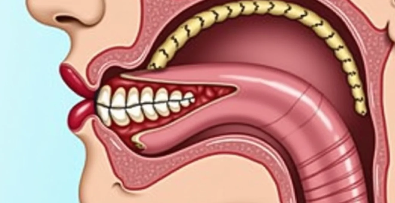
Thyroid disorders affecting the neck can create a peculiar and often distressing sensation that something is lodged in the throat. This phenomenon, medically termed globus pharyngeus when related to thyroid pathology, affects millions of individuals worldwide and represents one of the most common presentations in otolaryngological practice. The thyroid gland’s strategic anatomical position at the base of the neck means that even modest enlargement can exert significant pressure on surrounding structures, including the oesophagus, trachea, and recurrent laryngeal nerves.
Understanding the complex relationship between thyroid pathology and throat obstruction symptoms requires a comprehensive appreciation of cervical anatomy, pathophysiological mechanisms, and the diverse clinical presentations that can emerge. Whether caused by benign multinodular goitre, inflammatory thyroiditis, or more concerning malignant processes, thyroid-related throat obstruction demands prompt recognition and appropriate management to prevent potentially serious complications.
Understanding thyroid gland anatomy and its relationship to pharyngeal compression
The thyroid gland’s intimate relationship with critical neck structures creates a complex anatomical environment where pathological changes can rapidly translate into symptomatic compression. Located in the lower anterior neck, the thyroid consists of two lobes connected by an isthmus, normally weighing approximately 20-25 grams in healthy adults. The gland’s position directly anterior to the trachea and inferior to the thyroid cartilage means that enlargement in any direction can potentially compromise surrounding structures.
Thyroid cartilage positioning and laryngeal framework impact
The thyroid cartilage forms the prominent laryngeal framework, commonly known as the Adam’s apple, and serves as a crucial landmark for understanding thyroid pathology. When thyroid enlargement occurs, particularly in the superior poles of the lateral lobes, direct contact with the thyroid cartilage can create mechanical compression effects. This compression mechanism often manifests as voice changes, including hoarseness, vocal fatigue, and alterations in pitch control.
The relationship between thyroid pathology and laryngeal framework dysfunction extends beyond simple mechanical compression. Inflammatory processes within the thyroid can create local tissue oedema that affects the mobility of laryngeal cartilages, particularly during swallowing and phonation. Patients frequently describe a sensation of throat tightness that worsens during periods of vocal use or when attempting to clear the throat.
Recurrent laryngeal nerve pathway and vocal cord function
The recurrent laryngeal nerves follow a predictable anatomical course that places them in close proximity to the posterior aspect of the thyroid gland. These nerves, branches of the vagus nerve, innervate all intrinsic laryngeal muscles except the cricothyroid muscle. Understanding their pathway is crucial because thyroid pathology can affect nerve function through direct compression, inflammatory infiltration, or surgical trauma.
Benign thyroid enlargement typically causes gradual nerve compression, leading to subtle voice changes that patients may initially attribute to other causes. However, rapid thyroid expansion , whether from haemorrhage into a nodule or aggressive inflammatory processes, can create acute nerve compression with sudden onset vocal cord paralysis. The left recurrent laryngeal nerve is particularly vulnerable due to its longer course around the aortic arch.
Oesophageal compression mechanisms in thyroid enlargement
Oesophageal compression represents one of the most clinically significant complications of thyroid enlargement, particularly when posterior extension occurs. The oesophagus lies directly posterior to the trachea and thyroid gland, making it susceptible to compression when thyroid pathology extends into the retrotracheal space. This compression mechanism typically develops gradually, allowing for some compensatory adaptation, but can eventually lead to significant dysphagia.
The degree of oesophageal compression correlates strongly with the size and location of thyroid pathology. Retrosternal goitre extension creates particularly challenging compression patterns because the thoracic inlet provides limited space for accommodation. Patients with oesophageal compression often report difficulty swallowing solid foods initially, with progression to liquid dysphagia in severe cases.
Tracheal deviation patterns in multinodular goitre
Tracheal deviation occurs when thyroid enlargement becomes sufficiently large to displace the trachea from its normal midline position. This phenomenon is most commonly observed in longstanding multinodular goitre, where asymmetric growth patterns create unequal pressure distribution. The trachea typically deviates away from the side of greatest thyroid enlargement, though bilateral enlargement can cause anterior displacement without lateral deviation.
Understanding tracheal deviation patterns is crucial for surgical planning and risk assessment. Significant tracheal displacement can indicate the presence of retrosternal extension, which may require modified surgical approaches or specialised anaesthetic management. Cross-sectional imaging studies reveal that even modest tracheal deviation often represents more extensive thyroid pathology than suggested by physical examination alone.
Pathophysiology of Thyroid-Related globus pharyngeus syndrome
Thyroid-related globus pharyngeus syndrome represents a complex interplay of mechanical compression, inflammatory responses, and neurological dysfunction that creates the characteristic sensation of throat obstruction. This syndrome can develop through multiple pathophysiological mechanisms, each contributing to the overall symptom complex that patients experience. Understanding these mechanisms is essential for developing targeted treatment strategies and predicting patient outcomes.
The pathophysiology involves both direct mechanical effects and indirect neurological responses to thyroid pathology. Mechanical compression of pharyngeal and laryngeal structures creates immediate pressure sensations, while inflammatory mediators released from diseased thyroid tissue can sensitise local nerve endings and create heightened awareness of normal anatomical structures. This dual mechanism explains why some patients experience symptoms disproportionate to the apparent degree of thyroid enlargement on imaging studies.
Hashimoto’s thyroiditis inflammatory response and tissue swelling
Hashimoto’s thyroiditis creates a unique pathophysiological environment characterised by chronic lymphocytic infiltration and progressive tissue destruction. The inflammatory response generates significant local tissue oedema that can extend beyond the thyroid capsule to affect surrounding structures. This inflammatory process often creates symptoms before significant thyroid enlargement becomes apparent on physical examination or routine imaging.
The autoimmune nature of Hashimoto’s thyroiditis means that inflammatory mediators, including various cytokines and chemokines, circulate locally and can affect nerve sensitivity in the surrounding tissues. Patients with active thyroiditis frequently report fluctuating symptoms that correlate with inflammatory activity rather than thyroid size alone. This variability in symptom severity can create diagnostic challenges, particularly in early disease stages.
Papillary thyroid carcinoma infiltration patterns
Papillary thyroid carcinoma demonstrates characteristic infiltration patterns that can create throat obstruction symptoms through mechanisms distinct from benign thyroid enlargement. Unlike benign processes that typically respect tissue planes, malignant thyroid tissue can infiltrate surrounding structures, including the tracheal wall, oesophageal muscle, and recurrent laryngeal nerve pathways. This infiltrative behaviour creates more severe and progressive symptoms compared to benign pathology of similar size.
The biological behaviour of papillary thyroid carcinoma includes a tendency for local invasion that can affect critical structures even when the primary tumour remains relatively small. Extrathyroidal extension occurs in approximately 30% of papillary thyroid cancers and significantly increases the risk of developing throat obstruction symptoms. Early recognition of malignant infiltration patterns is crucial for appropriate surgical planning and patient counselling regarding potential postoperative complications.
Substernal goitre extension and thoracic inlet obstruction
Substernal goitre extension represents a unique pathophysiological challenge where thyroid tissue grows beyond the thoracic inlet into the superior mediastinum. This extension pattern creates compression effects that differ significantly from cervical thyroid pathology due to the rigid bony boundaries of the thoracic inlet. The confined space limits accommodation for expanding thyroid tissue, leading to more severe compression effects on the trachea and oesophagus.
The development of substernal extension typically occurs gradually over many years, allowing for compensatory changes in surrounding structures. However, acute complications can arise from haemorrhage into substernal components or rapid growth phases. Emergency presentations of substernal goitre can include acute airway obstruction, particularly when patients assume supine positioning or experience upper respiratory tract infections that increase airway resistance.
Thyroglossal duct cyst complications and airway compromise
Thyroglossal duct cysts represent developmental anomalies that can create throat obstruction through mechanisms related to their unique anatomical relationships. These cysts can occur anywhere along the path of thyroid descent during embryogenesis, from the base of the tongue to the thyroid gland itself. When located near the hyoid bone or in the suprahyoid region, infected or rapidly enlarging thyroglossal duct cysts can create significant airway compression.
The pathophysiology of thyroglossal duct cyst complications involves both mass effect and inflammatory responses. Cyst infection can lead to rapid expansion and surrounding cellulitis that compromises airway patency. The close relationship between thyroglossal duct cysts and the hyoid bone means that surgical management requires careful consideration of anatomical relationships to prevent recurrence while maintaining airway integrity.
Clinical manifestation patterns of Thyroid-Induced dysphagia
Thyroid-induced dysphagia presents through distinctive clinical patterns that reflect the specific anatomical relationships between enlarged thyroid tissue and surrounding structures. Patients typically develop a progressive constellation of symptoms that begin subtly and intensify as thyroid pathology advances. The clinical presentation often follows predictable patterns based on the location and nature of thyroid enlargement, making recognition possible even before advanced imaging studies are available.
Early symptoms commonly include a sensation of throat fullness or the feeling that something is lodged in the neck area. This globus sensation typically worsens during swallowing and may be accompanied by frequent throat clearing attempts. Patients often describe the sensation as similar to having a pill stuck in the throat or feeling as though they need to swallow repeatedly to clear an obstruction that never fully resolves. These initial symptoms can be intermittent and may fluctuate with factors such as stress, fatigue, or thyroid hormone fluctuations.
As thyroid enlargement progresses, mechanical compression symptoms become more pronounced and persistent. Solid food dysphagia typically develops before liquid swallowing difficulties, reflecting the progressive nature of oesophageal compression. Patients report increased difficulty with foods that require more coordinated swallowing mechanisms, such as bread, meat, or sticky foods. The development of liquid dysphagia suggests significant oesophageal compression and requires urgent evaluation to prevent aspiration complications.
Respiratory symptoms accompanying thyroid-related dysphagia include positional shortness of breath, particularly when lying flat or during physical exertion. The sensation of incomplete inspiration or the need to take frequent deep breaths often develops gradually and may be attributed to other causes before thyroid pathology is recognised. Nocturnal symptoms can include snoring, sleep disruption, or the sensation of choking during sleep, particularly when thyroid enlargement creates significant tracheal compression.
Voice changes represent another important component of the clinical presentation, ranging from subtle alterations in vocal quality to complete voice loss in severe cases. Patients may notice vocal fatigue during extended speaking, changes in pitch range, or a hoarse, breathy voice quality. Professional voice users, such as teachers or singers, often detect these changes earlier than the general population due to their heightened awareness of vocal function.
The clinical progression of thyroid-related throat obstruction typically follows a predictable timeline, with early globus sensations progressing to mechanical dysphagia and eventually respiratory compromise if left untreated. Recognition of this progression pattern enables healthcare providers to intervene before life-threatening complications develop.
Diagnostic imaging protocols for Thyroid-Associated throat obstruction
Comprehensive imaging evaluation of thyroid-associated throat obstruction requires a systematic approach that addresses both thyroid pathology and its effects on surrounding structures. Modern imaging protocols have evolved to provide detailed assessment of thyroid morphology, compression effects, and the extent of any retrosternal extension. The choice of imaging modality depends on the clinical presentation, suspected pathology, and the need to evaluate specific anatomical relationships.
High-resolution ultrasound assessment of thyroid nodule compression
High-resolution ultrasound serves as the primary imaging modality for initial thyroid assessment, providing detailed characterisation of thyroid morphology and nodular pathology. Modern ultrasound systems with high-frequency transducers can detect nodules as small as 2-3 millimetres and assess their relationship to surrounding structures. The real-time nature of ultrasound examination allows for dynamic assessment of thyroid movement during swallowing and evaluation of compression effects on adjacent structures.
Ultrasound assessment of compression effects focuses on evaluating tracheal position, oesophageal displacement, and the relationship between thyroid pathology and the carotid sheaths. Colour Doppler imaging can assess vascular displacement and identify any compromise of major neck vessels. The examination should include assessment of both thyroid lobes, the isthmus, and any visible retrosternal extension, though ultrasound has limitations in evaluating substernal components due to acoustic shadowing from overlying bone and air.
CT angiography evaluation of retrosternal goitre extension
Computed tomography with intravenous contrast enhancement provides superior evaluation of retrosternal goitre extension and its relationship to mediastinal structures. CT angiography protocols specifically designed for thyroid evaluation include thin-section imaging through the neck and upper chest with multiplanar reconstruction capabilities. This imaging approach allows precise measurement of tracheal diameter, assessment of oesophageal displacement, and evaluation of the relationship between thyroid tissue and major vascular structures.
The ability to assess the degree of tracheal narrowing quantitatively makes CT particularly valuable for surgical planning and risk stratification. Cross-sectional area measurements of the trachea at the level of maximum compression provide objective data for determining the urgency of surgical intervention. CT also excels at identifying calcifications within thyroid nodules, which may indicate specific pathological processes or increase surgical complexity.
Barium swallow studies for oesophageal displacement documentation
Barium swallow studies provide dynamic functional assessment of swallowing mechanics and can demonstrate the degree of oesophageal compression and displacement. These studies are particularly valuable when patients report significant dysphagia symptoms that seem disproportionate to the apparent degree of thyroid enlargement on cross-sectional imaging. The real-time visualisation of barium passage through the oesophagus can reveal functional obstruction that may not be apparent on static imaging studies.
Modified barium swallow studies can assess both liquid and solid bolus passage, providing information about the functional impact of thyroid-related compression. Video fluoroscopic evaluation allows frame-by-frame analysis of swallowing mechanics and can identify subtle coordination abnormalities that contribute to patient symptoms. These studies are particularly useful for documenting baseline function before surgical intervention and monitoring improvement following treatment.
MRI protocol for soft tissue invasion assessment
Magnetic resonance imaging provides superior soft tissue contrast resolution compared to CT, making it particularly valuable for assessing potential malignant invasion of surrounding structures. MRI protocols for thyroid evaluation typically include T1-weighted, T2-weighted, and post-contrast sequences in multiple planes. The excellent soft tissue differentiation capabilities of MRI allow detailed assessment of thyroid capsule integrity and evaluation of possible extrathyroidal extension.
The ability to assess tissue characteristics using various MRI sequences can help differentiate between benign and malignant thyroid pathology. Diffusion-weighted imaging sequences can provide additional information about tissue cellularity and may help identify areas of particular concern for malignancy. MRI is particularly valuable when CT findings suggest possible invasion of critical structures such as the recurrent laryngeal nerve pathways or major blood vessels.
Endocrine function testing in compressive thyroid disorders
Endocrine function assessment plays a crucial role in evaluating patients with compressive thyroid disorders, as thyroid hormone status can significantly influence symptom severity and treatment planning. The relationship between thyroid hormone levels and compression symptoms is complex, with both hyperthyroid and hypothyroid states potentially exacerbating throat obstruction sensations through different mechanisms. Comprehensive endocrine evaluation should include assessment of thyroid hormone levels, thyroid-stimulating hormone, and relevant antibody studies.
Thyroid-stimulating hormone (
TSH) represents the primary screening test for thyroid dysfunction, as it provides sensitive detection of both hyperthyroid and hypothyroid states. In patients with compressive thyroid symptoms, TSH levels help determine whether hormonal abnormalities contribute to symptom severity or influence treatment decisions. Suppressed TSH levels in the context of thyroid enlargement may indicate autonomous thyroid function, which can complicate surgical planning and anaesthetic management.
Free thyroxine (T4) and triiodothyronine (T3) measurements provide direct assessment of circulating thyroid hormone levels and help characterise the degree of thyroid dysfunction. Patients with compressive symptoms may demonstrate normal thyroid hormone levels despite significant glandular enlargement, particularly in cases of multinodular goitre with preserved overall function. However, subclinical hyperthyroidism can exacerbate compression symptoms by increasing tissue metabolic activity and local blood flow.
Thyroid autoantibody testing, including thyroid peroxidase antibodies and thyroglobulin antibodies, helps identify autoimmune thyroid conditions that may influence treatment approaches. Hashimoto’s thyroiditis with positive antibodies often requires different surgical considerations compared to non-autoimmune thyroid enlargement. Thyroid-stimulating immunoglobulin testing may be indicated when Graves’ disease is suspected, as this condition can cause rapid thyroid enlargement with associated compression symptoms.
Surgical intervention strategies for thyroid-related airway compression
Surgical management of thyroid-related airway compression requires careful consideration of patient factors, degree of compression, and underlying thyroid pathology. The primary goals include relief of compression symptoms, restoration of normal anatomy, and prevention of recurrent disease while minimising surgical morbidity. Modern surgical approaches have evolved to incorporate minimally invasive techniques where appropriate, though traditional open approaches remain necessary for complex cases with significant retrosternal extension or suspected malignancy.
Patient selection for surgical intervention depends on symptom severity, imaging findings, and response to conservative management. Absolute indications for surgery include severe airway compromise, progressive dysphagia with nutritional impact, and suspected malignancy. Relative indications encompass cosmetic concerns, mild compression symptoms affecting quality of life, and thyroid dysfunction refractory to medical management. The decision-making process must balance symptom relief against surgical risks, particularly in elderly patients or those with significant comorbidities.
Total thyroidectomy techniques for massive goitre removal
Total thyroidectomy represents the gold standard approach for massive goitre causing significant compression symptoms, particularly when bilateral thyroid pathology exists or when retrosternal extension is present. The procedure requires meticulous technique to identify and preserve critical structures, including the recurrent laryngeal nerves and parathyroid glands. Preoperative planning with cross-sectional imaging helps anticipate surgical challenges and guide the optimal approach.
The surgical technique begins with careful dissection of the thyroid capsule to identify anatomical planes that may be distorted by longstanding enlargement. Nerve monitoring technology has become increasingly valuable in these challenging cases, providing real-time feedback about nerve function during dissection. The superior pole dissection requires particular attention to avoid injury to the external branch of the superior laryngeal nerve, which controls vocal pitch and projection.
Management of retrosternal components often requires finger dissection and careful mobilisation to deliver thyroid tissue into the neck incision. In cases where blind finger dissection proves inadequate, sternotomy may be necessary to achieve safe removal. The decision for sternotomy should be made early in the procedure rather than after prolonged unsuccessful attempts at cervical delivery, which can increase morbidity significantly.
Hemithyroidectomy approaches for unilateral nodular disease
Hemithyroidectomy offers an excellent option for patients with unilateral thyroid pathology causing compression symptoms, allowing preservation of thyroid function while addressing the symptomatic disease. This approach is particularly suitable for benign unilateral multinodular disease or large solitary nodules causing tracheal displacement or oesophageal compression. The preservation of contralateral thyroid tissue often eliminates the need for lifelong thyroid hormone replacement therapy.
The surgical technique for hemithyroidectomy follows similar principles to total thyroidectomy but focuses on complete removal of the affected lobe while preserving the isthmus and contralateral lobe when possible. Careful assessment of the contralateral lobe during surgery helps ensure that remaining thyroid tissue appears normal and capable of maintaining adequate hormone production. Intraoperative frozen section analysis may be valuable when nodular disease exists in the remaining lobe.
Postoperative monitoring following hemithyroidectomy includes serial assessment of thyroid function, as approximately 15-20% of patients may develop hypothyroidism requiring hormone replacement. The development of contralateral thyroid pathology remains a long-term consideration, necessitating ongoing surveillance with periodic ultrasound examinations and clinical evaluation.
Tracheostomy considerations in emergency airway management
Emergency airway management in patients with severe thyroid-related airway compression requires careful consideration of tracheostomy as a stabilising procedure before definitive thyroid surgery. Acute airway compromise can develop suddenly in patients with large goitres, particularly following upper respiratory infections, trauma, or haemorrhage into thyroid nodules. The decision to perform emergency tracheostomy versus proceeding directly to thyroidectomy depends on the severity of airway compromise and surgical expertise available.
Tracheostomy in the setting of thyroid enlargement presents unique technical challenges due to anatomical distortion and the potential for haemorrhage from enlarged thyroid vessels. The tracheostomy site may need to be positioned lower than usual due to thyroid enlargement, and careful dissection is required to avoid injury to enlarged thyroid vessels or aberrant thyroid tissue. Coordination between anaesthesia and surgical teams is crucial for managing the transition from endotracheal intubation to tracheostomy.
Following emergency tracheostomy, patients typically require prompt thyroid surgery once their condition stabilises. The presence of a tracheostomy can facilitate safer thyroidectomy by securing the airway and allowing for controlled ventilation throughout the procedure. However, the tracheostomy site must be carefully managed during thyroid surgery to prevent complications such as pneumothorax or subcutaneous emphysema.
Minimally invasive thyroidectomy protocols and patient selection
Minimally invasive thyroidectomy techniques have emerged as viable options for selected patients with thyroid-related compression symptoms, offering potential benefits including reduced scarring, decreased postoperative pain, and faster recovery times. However, patient selection criteria must be carefully applied to ensure safety and efficacy, as these techniques have limitations in managing large goitres or extensive retrosternal disease.
Endoscopic thyroidectomy approaches utilise small incisions placed away from the central neck, often in the axilla or through a transoral route. Patient selection criteria for minimally invasive approaches include thyroid size limitations, absence of significant retrosternal extension, and no evidence of malignancy with extrathyroidal extension. The learning curve for these techniques is substantial, requiring dedicated training and experience to achieve outcomes comparable to conventional surgery.
Robotic thyroidectomy represents the most advanced minimally invasive approach, offering enhanced visualisation and precision in instrument manipulation. The robotic platform provides three-dimensional visualisation and tremor filtration, potentially improving outcomes in complex cases. However, the increased operative time and cost associated with robotic surgery must be balanced against patient benefits, and long-term outcome data continues to accumulate to define optimal patient selection criteria.