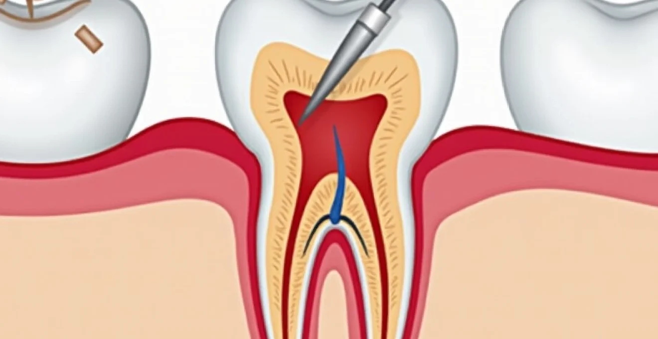
Post-extraction ulcers represent one of the most concerning complications patients face following dental surgery, affecting up to 15% of individuals who undergo tooth removal procedures. These painful lesions can significantly impact quality of life, hindering normal eating, speaking, and oral hygiene practices during the critical healing period. Understanding the complex interplay between tissue trauma, bacterial colonisation, and individual healing responses is essential for both preventing and managing these troublesome complications effectively.
The development of ulcers following tooth extraction stems from a multifaceted process involving disrupted tissue architecture, compromised vascular supply, and altered oral microflora. Modern dental practice has evolved to recognise these ulcerative complications as preventable conditions, with evidence-based protocols now available to minimise risk and optimise healing outcomes. The distinction between normal healing discomfort and pathological ulceration requires careful assessment and timely intervention to prevent prolonged morbidity.
Post-extraction socket healing physiology and ulceration mechanisms
The normal healing process following tooth extraction involves a precisely orchestrated sequence of biological events that can be disrupted at multiple stages. Understanding this physiological cascade provides crucial insights into why certain individuals develop ulcerative complications whilst others experience uncomplicated recovery.
Alveolar bone remodelling after tooth removal
Alveolar bone remodelling begins immediately following tooth extraction, with osteoclastic activity initiating within 24-48 hours of the procedure. This bone remodelling process involves the systematic breakdown of the lamina dura and surrounding cortical bone, creating an environment where bacterial infiltration can occur more readily. The exposed bone surface becomes vulnerable to infection and subsequent ulceration, particularly in cases where the overlying soft tissue fails to achieve adequate coverage.
The remodelling phase typically extends over 3-6 months, during which time the alveolar ridge undergoes significant dimensional changes. Approximately 40-60% of the original bone width is lost during this period, with the greatest changes occurring within the first three months. This extensive bone turnover creates pockets and irregularities that can trap food debris and bacteria, predisposing the area to ulcerative breakdown.
Epithelial migration and soft tissue closure timeline
Epithelial migration across the extraction socket represents a critical phase in preventing ulcer formation. Under optimal conditions, epithelial cells begin migrating from the wound margins within 24-48 hours, achieving complete surface coverage within 7-14 days. However, this process can be significantly delayed by factors such as smoking, poor oral hygiene, or systemic conditions affecting wound healing.
The quality of epithelial closure directly correlates with ulcer risk, as incomplete or fragile epithelial coverage leaves the underlying connective tissue exposed to mechanical trauma and bacterial colonisation. Studies demonstrate that delayed epithelial migration occurs in approximately 20% of extraction cases, particularly following surgical removal of impacted teeth or in patients with compromised healing capacity.
Bacterial colonisation patterns in fresh extraction sites
The extraction socket presents an ideal environment for bacterial colonisation, with the blood clot providing nutrients and the disrupted tissue architecture offering reduced immune surveillance. Within 6-12 hours post-extraction, bacterial counts can increase 100-fold compared to intact oral tissues, with anaerobic species predominating in the deeper aspects of the socket.
Streptococcus species and Actinomyces typically establish early colonisation, followed by more pathogenic organisms such as Porphyromonas gingivalis and Fusobacterium nucleatum . This bacterial succession creates conditions conducive to ulcer formation, particularly when combined with mechanical irritation or compromised host defences. The biofilm formation that occurs within 48-72 hours further protects these organisms from antimicrobial therapy and immune clearance mechanisms.
Inflammatory response cascade and cytokine release
The inflammatory response following tooth extraction involves complex cytokine networks that regulate both healing and tissue destruction. Pro-inflammatory mediators such as interleukin-1β, tumour necrosis factor-α, and prostaglandin E2 reach peak levels within 24-48 hours post-extraction, creating an environment where excessive inflammation can impede normal healing processes.
Dysregulated cytokine production, particularly prolonged elevation of matrix metalloproteinases, can lead to excessive tissue breakdown and ulcer formation. This biochemical imbalance is more common in patients with systemic inflammatory conditions, diabetes mellitus, or those taking immunosuppressive medications. The balance between pro-inflammatory and anti-inflammatory signals determines whether healing proceeds normally or progresses to chronic ulceration.
Primary aetiological factors contributing to Post-Extraction ulcer formation
Multiple interconnected factors contribute to ulcer development following tooth extraction, ranging from surgical technique variables to patient-specific risk factors. Recognition of these aetiological elements enables targeted prevention strategies and early intervention protocols.
Traumatic extraction techniques and tissue damage assessment
Excessive force during tooth extraction represents the most significant modifiable risk factor for subsequent ulcer formation. Traumatic extraction techniques that involve extensive bone removal, aggressive tissue elevation, or prolonged procedure times create larger wounds with compromised vascular supply. The degree of initial tissue trauma directly correlates with healing complications, including ulcer development.
Assessment of extraction complexity using standardised scoring systems helps predict ulcer risk and guide appropriate surgical approaches. Simple extractions with intact crown structure and adequate periodontal support carry minimal ulcer risk, whilst surgical extractions requiring bone removal or tooth sectioning increase complication rates by 300-400%. The use of minimally invasive extraction techniques, such as periotome-assisted removal or piezosurgery, significantly reduces tissue trauma and subsequent ulceration risk.
Compromised local blood supply and ischaemic ulceration
Adequate vascularisation is essential for normal wound healing and ulcer prevention. Local factors that compromise blood supply, such as excessive vasoconstrictor use in local anaesthetic solutions, aggressive tissue handling, or tight suturing, create ischaemic conditions predisposing to ulcer formation. The posterior mandible and maxillary molar regions are particularly vulnerable due to their naturally reduced vascular density.
Ischaemic ulceration typically develops 3-7 days post-extraction and presents with characteristic pale, necrotic tissue surrounded by an inflammatory border. These ulcers are often deeper and more painful than traumatic ulcers, requiring specific management approaches including debridement and antimicrobial therapy. Smoking cessation is crucial, as tobacco use reduces local blood flow by up to 50% and significantly impairs healing capacity.
Xerostomia-induced delayed wound healing complications
Xerostomia, or dry mouth, affects approximately 20% of adults and represents a significant risk factor for post-extraction ulceration. Reduced salivary flow compromises the oral cavity’s natural cleansing mechanisms and reduces the delivery of growth factors and antimicrobial proteins essential for healing. Patients with xerostomia demonstrate 2-3 times higher rates of extraction site complications compared to those with normal salivary function.
The impact of xerostomia extends beyond simple moisture reduction, as saliva contains numerous healing-promoting factors including epidermal growth factor, transforming growth factor-β, and immunoglobulin A. Without adequate salivary protection, extraction sites become vulnerable to desiccation, bacterial overgrowth, and mechanical trauma from food particles. Management strategies must address both symptom relief and underlying causes of salivary dysfunction.
Immunosuppressive medications and ulcer risk stratification
Patients taking immunosuppressive medications face substantially elevated risks of post-extraction ulceration due to impaired wound healing and increased infection susceptibility. Corticosteroids , methotrexate, and biological therapies can delay healing by 50-100% and increase ulcer formation rates by 400-500%. The timing of dental extractions in relation to immunosuppressive therapy requires careful coordination with prescribing physicians.
Risk stratification protocols should categorise patients based on medication type, dosage, and duration of therapy, with high-risk individuals receiving prophylactic antimicrobial coverage and enhanced monitoring protocols.
Differential diagnosis of Post-Extraction oral ulcerations
Accurate differential diagnosis of post-extraction ulcerations is crucial for appropriate management, as various conditions can present with similar clinical features but require distinctly different treatment approaches. The timing, location, and morphological characteristics of ulcers provide essential diagnostic clues.
Aphthous stomatitis versus traumatic ulceration clinical markers
Distinguishing between aphthous stomatitis and traumatic ulceration requires careful assessment of ulcer characteristics, timing, and patient history. Aphthous ulcers typically present with well-defined borders, a characteristic yellow-white fibrinous base, and an erythematous halo. These lesions often develop 2-5 days post-extraction and may occur in multiple sites throughout the oral cavity, suggesting a systemic rather than localised aetiology.
Traumatic ulcerations, in contrast, directly correlate with the extraction site and demonstrate irregular borders consistent with the mechanism of injury. The timing of traumatic ulcers is typically immediate or within 24 hours of the procedure, and they rarely occur in locations distant from the surgical site. Pain intensity tends to be greater with aphthous ulcers, whilst traumatic ulcers often respond more readily to topical anaesthetic agents.
Osteomyelitis-related ulceration and radiographic findings
Osteomyelitis of the extraction socket represents a serious complication that can present with persistent ulceration and systemic symptoms. Radiographic evidence of bone destruction, including loss of lamina dura continuity, irregular bone margins, or sequestrum formation, distinguishes osteomyelitis from simple ulceration. These changes typically become apparent 10-14 days after the initial procedure, though clinical symptoms may precede radiographic findings.
The presence of purulent discharge, mobile bone fragments, or expanding areas of bone exposure strongly suggests osteomyelitic involvement. Panoramic radiography provides initial screening, but cone-beam computed tomography offers superior detail for assessing bone destruction extent and treatment planning. Early recognition and aggressive management are essential to prevent progression to chronic osteomyelitis or pathological fracture.
Malignant transformation signs in persistent extraction site ulcers
Persistent ulceration beyond 3-4 weeks raises concerns for potential malignant transformation or pre-existing undiagnosed malignancy. Squamous cell carcinoma can masquerade as chronic extraction site ulceration, particularly in patients with tobacco or alcohol use histories. Suspicious features include indurated borders, rapid expansion, painless progression, or involvement of adjacent anatomical structures.
The concept of field cancerisation suggests that apparently normal tissue adjacent to extraction sites may harbour premalignant changes, predisposing to malignant transformation following tissue trauma. Any ulcer demonstrating atypical features or failing to respond to conventional therapy within 2-3 weeks warrants tissue biopsy for histopathological examination. Early detection significantly improves treatment outcomes and long-term prognosis.
Bisphosphonate-related osteonecrosis of the jaw presentation
Bisphosphonate-related osteonecrosis of the jaw (BRONJ) represents a devastating complication that can present as non-healing extraction site ulceration with exposed bone. The condition affects 1-15% of patients taking bisphosphonates, with higher risks associated with intravenous formulations and longer treatment durations. BRONJ typically develops 2-6 months post-extraction but can occur years after bisphosphonate discontinuation.
Clinical presentation includes persistent bone exposure, purulent discharge, and progressive pain. The exposed bone appears yellow-white and demonstrates a rough, irregular surface texture. Radiographic findings may be minimal initially but can progress to show extensive bone sclerosis, sequestrum formation, and pathological fracture. Prevention through pre-extraction risk assessment and alternative treatment planning remains the most effective management strategy.
Current guidelines recommend bone-sparing procedures whenever possible in bisphosphonate-exposed patients, with extraction reserved for cases where conservative management has failed.
Evidence-based therapeutic interventions for extraction site ulcers
Contemporary management of post-extraction ulcers relies on evidence-based protocols that address both symptomatic relief and underlying pathophysiology. Treatment selection depends on ulcer aetiology, severity, and patient-specific factors, with multimodal approaches demonstrating superior outcomes compared to single-intervention strategies.
Topical therapies form the cornerstone of ulcer management, with corticosteroid preparations showing particular efficacy for inflammatory ulcerations. Triamcinolone acetonide 0.1% in orabase demonstrates significant pain reduction and accelerated healing when applied 3-4 times daily. However, prolonged steroid use can impair healing and increase infection risk, necessitating careful monitoring and time-limited treatment courses.
Antimicrobial interventions play a crucial role in managing infected ulcerations and preventing secondary bacterial complications. Chlorhexidine gluconate 0.12% rinses provide broad-spectrum antimicrobial activity whilst maintaining compatibility with healing tissues. For severe infections, systemic antibiotic therapy targeting oral anaerobes may be necessary, with amoxicillin-clavulanate or metronidazole representing first-line choices in the absence of specific contraindications.
Advanced therapeutic modalities including low-level laser therapy, platelet-rich plasma application, and tissue-engineered scaffolds show promise for managing refractory ulcerations. Photobiomodulation therapy , increasingly available at contemporary dental facilities in Montreal, uses 660-830nm wavelengths to achieve significant anti-inflammatory and cellular proliferative effects. Treatment protocols typically involve 3-5 sessions over 1-2 weeks, with each session lasting 2-5 minutes depending on specific ulcer characteristics.
Pain management requires a multimodal approach combining topical anaesthetics, systemic analgesics, and non-pharmacological interventions. Benzocaine 20% gels provide rapid onset topical anaesthesia, though caution is warranted regarding methemoglobinemia risk with excessive use. Systemic analgesics should follow the WHO pain ladder, beginning with paracetamol and progressing through NSAIDs to opioid preparations for severe pain. The addition of gabapentin or pregabalin may benefit patients with neuropathic pain components.
Pharmaceutical management protocols and antimicrobial selection
Rational pharmaceutical management of post-extraction ulcers requires understanding of local tissue pharmacokinetics, bacterial resistance patterns, and potential drug interactions. The selection of appropriate antimicrobial agents must balance efficacy against target pathogens with minimal disruption to normal healing processes and commensal oral flora.
Topical antimicrobial agents offer the advantage of high local concentrations with minimal systemic exposure, making them ideal for localised ulcerative lesions. Metronidazole gel 0.75% demonstrates excellent activity against anaerobic pathogens commonly associated with extraction site infections whilst avoiding the broad-spectrum effects that can predispose to candidal overgrowth. Application frequency of 2-3 times daily provides sustained antimicrobial activity throughout the healing period.
Systemic antibiotic selection should be guided by local resistance patterns and patient-specific factors including allergies, renal function, and concurrent medications. The oral microflora’s complex ecology requires agents with appropriate spectra that target pathogenic organisms whilst preserving beneficial species. Amoxicillin 500mg three times daily remains highly effective against streptococcal species, whilst the addition of clavulanic acid extends coverage to β-lactamase-producing organisms.
For patients with β-lactam allergies, alternative regimens including doxycycline 100mg twice daily or azithromycin 500mg daily provide excellent oral tissue penetration and anti-inflammatory properties beyond their antimicrobial effects. The extended half-life of azithromycin allows once-daily dosing, improving compliance in outpatient management. However, the increasing prevalence of macrolide resistance among oral streptococci necessitates careful monitoring and consideration of alternative agents in treatment failures.
Antifungal prophylaxis should be considered in patients receiving broad-spectrum antibiotics for extended periods,
particularly those with diabetes or immunocompromise. Nystatin oral suspension 100,000 units/mL administered four times daily provides effective coverage against Candida species without significant drug interactions. The concurrent use of probiotics containing Lactobacillus reuteri can help maintain oral microbiome balance during antimicrobial therapy, reducing the risk of opportunistic infections and supporting natural healing mechanisms.
Corticosteroid therapy requires careful risk-benefit assessment, as these agents can simultaneously reduce inflammation and impair immune function. Topical preparations offer superior safety profiles compared to systemic administration, with triamcinolone acetonide 0.1% providing excellent anti-inflammatory activity when applied directly to ulcerated tissues. Treatment duration should be limited to 7-10 days to minimise adverse effects, with gradual tapering to prevent rebound inflammation.
The emergence of antimicrobial resistance necessitates judicious use of antibiotics and consideration of alternative therapeutic approaches. Bacteriophage therapy represents a promising future direction, offering highly specific targeting of pathogenic bacteria without affecting beneficial commensals. Current research demonstrates efficacy against biofilm-associated infections, which are particularly relevant in the complex microbial environment of extraction sites.
Prevention strategies and pre-extraction risk assessment protocols
Comprehensive prevention strategies begin with thorough pre-extraction risk assessment and continue through optimised surgical technique and post-operative care protocols. Evidence-based prevention approaches can reduce ulcer incidence by up to 70%, making systematic implementation of these strategies a clinical imperative for all practitioners performing dental extractions.
Pre-operative risk stratification utilises validated scoring systems that incorporate patient demographics, medical history, medication use, and local tissue factors. High-risk patients include those over 65 years, diabetics with HbA1c >7%, smokers consuming >10 cigarettes daily, and individuals taking immunosuppressive medications. Bisphosphonate exposure history requires particular attention, with dental management guidelines recommending drug holidays where medically appropriate before elective extractions.
Optimised surgical technique represents the most significant modifiable factor in ulcer prevention. Minimally invasive extraction protocols emphasising tissue preservation, gentle handling, and adequate haemostasis reduce tissue trauma by 60-80% compared to conventional approaches. The use of periotomes for periodontal ligament severing, controlled force application, and immediate socket preservation techniques all contribute to improved healing outcomes and reduced ulcer formation.
Post-operative instructions must be tailored to individual risk profiles, with high-risk patients receiving enhanced monitoring and modified care protocols. Standard instructions include avoiding vigorous rinsing for 24 hours, maintaining soft diet consistency for 48-72 hours, and implementing gentle oral hygiene measures around the extraction site. Smoking cessation counselling should be provided to all tobacco users, with nicotine replacement therapy offered where appropriate to support cessation efforts during the critical healing period.
The implementation of chlorhexidine rinses beginning 24 hours post-extraction has demonstrated significant reductions in bacterial colonisation and subsequent infection rates. A 0.12% solution used twice daily for 7-10 days provides optimal antimicrobial activity whilst minimising taste alteration and staining effects. Patient education regarding proper rinsing technique ensures therapeutic concentrations reach the extraction site without disrupting blood clot formation.
Prevention protocols should be standardised across practice settings, with regular audit and quality improvement initiatives ensuring consistent implementation and optimal patient outcomes.
Nutritional optimisation plays a crucial role in prevention, particularly for patients with identified deficiencies or compromised healing capacity. Vitamin C supplementation at 500mg twice daily supports collagen synthesis and immune function, whilst zinc supplementation at 15mg daily enhances epithelial healing and reduces infection risk. Patients with diabetes require careful glucose management during the perioperative period, with target blood glucose levels maintained below 180mg/dL to optimise healing conditions.
Follow-up scheduling should reflect individual risk profiles, with high-risk patients receiving review appointments within 48-72 hours and standard-risk patients at 7-10 days post-extraction. Early detection of healing complications allows for prompt intervention and significantly improved outcomes. Telemedicine platforms can supplement in-person reviews, enabling patients to share photographs of healing sites and receive guidance on symptom management between appointments.
The integration of patient-reported outcome measures (PROMs) into follow-up protocols provides valuable insights into healing quality and patient experience. Simple visual analogue scales for pain, function, and satisfaction can identify patients requiring additional support or intervention. This data also supports continuous quality improvement initiatives and helps refine prevention protocols based on real-world effectiveness.