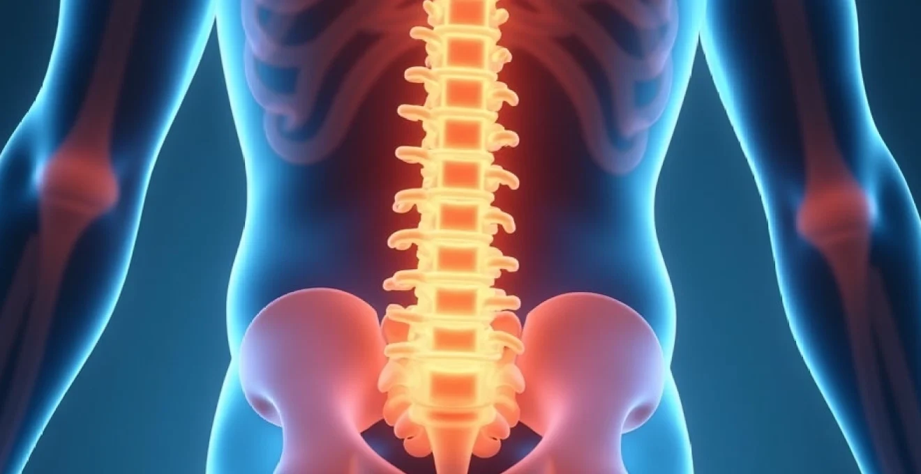
An L5-S1 annular fissure represents one of the most clinically significant spinal pathologies affecting the lumbosacral junction, where the fifth lumbar vertebra meets the first sacral vertebra. This critical anatomical region bears substantial mechanical loads and experiences considerable stress during daily activities, making it particularly vulnerable to disc degeneration and annular tears. The symptoms arising from L5-S1 annular fissures can be remarkably diverse, ranging from subtle lower back discomfort to debilitating radicular pain that significantly impacts quality of life. Understanding the complex symptomatology of these lesions requires a comprehensive appreciation of the intricate anatomical relationships, biomechanical forces, and neurological pathways involved in this region. The manifestations of L5-S1 annular fissures often present diagnostic challenges for healthcare practitioners, as symptoms can mimic other spinal conditions whilst varying considerably in their severity and distribution patterns.
Anatomical structure and pathophysiology of L5-S1 annular fissures
Annulus fibrosus composition and laminar organisation at L5-S1 junction
The annulus fibrosus at the L5-S1 level exhibits a unique structural composition that predisposes this region to specific patterns of failure and symptom development. Comprising approximately 15-20 concentric lamellae of type I collagen fibres, the annulus demonstrates alternating fibre orientations that typically range between 30-65 degrees relative to the horizontal plane. This sophisticated arrangement provides the disc with its remarkable tensile strength and ability to resist multidirectional loads. However, the posterolateral aspect of the L5-S1 annulus contains a higher proportion of vertically oriented fibres, creating a relative weak point that becomes particularly susceptible to tear formation during combined flexion and rotation movements.
The transition zone between the nucleus pulposus and annulus fibrosus at L5-S1 represents another critical area where fissures commonly initiate. This region experiences significant hydrostatic pressure variations throughout the day, with pressures reaching up to 2.3 times body weight during sitting activities. The laminar structure becomes progressively less organised towards the nucleus, and the collagen content decreases whilst proteoglycan concentration increases. When annular fissures develop in this transitional zone, they can propagate both radially towards the disc periphery and circumferentially around the nucleus, creating complex tear patterns that influence symptom presentation and treatment response.
Biomechanical stress distribution in lumbosacral transitional anatomy
The L5-S1 segment experiences unique biomechanical stresses due to its position as the transitional zone between the mobile lumbar spine and the rigid sacral complex. During forward flexion, the posterior annulus experiences tensile stresses that can exceed 4.5 MPa, particularly in the posterolateral regions where annular fissures most commonly occur. The lumbosacral angle, typically ranging between 26-57 degrees in healthy individuals, creates a natural shear stress concentration at this level that increases significantly with disc degeneration. These mechanical factors explain why L5-S1 annular fissures often present with symptoms that worsen during flexion-based activities and improve with extension positioning.
The facet joint orientation at L5-S1 differs markedly from the upper lumbar levels, with the joints positioned more coronally to resist the increased shear forces at this level. However, this anatomical adaptation can create altered load distribution patterns when annular integrity is compromised. Research demonstrates that even small annular defects can increase facet joint loading by up to 70%, contributing to the development of secondary symptoms including facetogenic pain and accelerated arthritic changes. The complex interplay between disc pathology and facet joint mechanics helps explain the multifaceted nature of L5-S1 annular fissure symptoms.
Inflammatory cascade following annular tear formation
When an annular fissure develops at the L5-S1 level, it triggers a sophisticated inflammatory response that significantly contributes to symptom development. The tear allows nucleus pulposus material, rich in inflammatory mediators such as phospholipase A2, interleukin-1β, and tumour necrosis factor-α, to migrate into the highly innervated outer annular layers. This exposure creates an autoimmune-like reaction, as the nucleus pulposus is normally isolated from the immune system during embryonic development. The resulting inflammatory cascade can persist for weeks or months, explaining the chronic nature of many L5-S1 annular fissure symptoms.
The inflammatory process extends beyond the disc itself, affecting surrounding neural structures including the dorsal root ganglion, nerve roots, and epidural space. Inflammatory mediators can increase neural membrane permeability, reduce nerve conduction velocity, and lower the threshold for nociceptive activation. This biochemical sensitisation helps explain why patients with L5-S1 annular fissures often experience symptoms that seem disproportionate to the apparent structural damage visible on imaging studies. The inflammation can also affect local blood flow and tissue oxygenation, contributing to the development of chronic pain patterns and delayed healing responses.
Neural innervation patterns of the posterior longitudinal ligament
The posterior longitudinal ligament (PLL) and outer annular fibres at the L5-S1 level receive dense innervation from the sinuvertebral nerves, which originate from the spinal nerve roots and sympathetic trunk. This innervation pattern is particularly concentrated in the posterolateral regions where annular fissures most commonly occur. The sinuvertebral nerves contain both sensory and autonomic fibres, explaining why L5-S1 annular fissures can produce not only pain but also autonomic symptoms such as altered bowel or bladder function in severe cases. The nerve supply demonstrates significant anatomical variations between individuals, contributing to the variable symptom patterns observed clinically.
Recent neuroanatomical studies have identified the presence of substance P and calcitonin gene-related peptide (CGRP) containing nerve fibres within the outer annulus, particularly at lumbosacral levels. These neuropeptides play crucial roles in pain transmission and neurogenic inflammation, helping to explain the intense, burning quality of pain that many patients experience with L5-S1 annular fissures. The distribution of these specialised nerve fibres correlates closely with the typical pain referral patterns observed in clinical practice, extending from the lower back into the buttock, posterior thigh, and sometimes reaching the lateral leg and foot.
Primary neurological manifestations of L5-S1 disc fissuring
Radicular pain distribution along S1 dermatome pathway
The most characteristic neurological manifestation of L5-S1 annular fissures involves radicular pain following the S1 dermatome distribution. This pain typically originates in the lower back, often described as a deep, aching sensation that can intensify to sharp, shooting discomfort. The pain characteristically radiates through the buttock, following the posterior and lateral aspects of the thigh before continuing down the posterolateral leg to the lateral border of the foot and little toe. The intensity and exact distribution can vary considerably between individuals, with some patients experiencing the full dermatomal pattern whilst others may have more limited referral zones.
The quality of S1 radicular pain often differs from mechanical back pain, presenting as a burning, electric, or stabbing sensation that patients frequently describe as unlike any pain they have previously experienced. This distinctive character results from the direct irritation of nerve root fibres by inflammatory mediators leaking from the fissured disc. The pain may be constant or intermittent, with many patients reporting episodic exacerbations that correlate with specific activities or positions. Night pain is common, often disrupting sleep patterns and contributing to the significant functional impact of L5-S1 annular fissures on daily life.
Motor weakness in plantarflexion and gastrocnemius function
Motor deficits associated with L5-S1 annular fissures primarily affect muscles innervated by the S1 nerve root, most notably the gastrocnemius-soleus complex responsible for plantarflexion. Patients may notice difficulty with activities requiring strong plantarflexion such as walking on toes, climbing stairs, or pushing off during running or jumping activities. The weakness typically develops gradually and may be subtle in the early stages, often becoming apparent only during specific functional assessments or when comparing strength between the affected and unaffected sides.
Clinical testing reveals weakness in plantarflexion that may be accompanied by atrophy of the calf muscles in chronic cases. The gastrocnemius muscle, being the largest component of the posterior compartment, shows the most obvious changes, with patients sometimes noting a visible difference in calf size between legs. This motor weakness can significantly impact functional activities, particularly those requiring propulsion or balance control. The degree of weakness generally correlates with the severity of nerve root compromise, with complete tears or large fissures producing more pronounced deficits than smaller, contained tears.
Sensory deficits affecting lateral foot and little toe sensation
Sensory disturbances represent another hallmark of L5-S1 annular fissure pathology, typically manifesting as altered sensation along the S1 dermatome. Patients commonly report numbness, tingling, or a “pins and needles” sensation affecting the lateral aspect of the foot and the fourth and fifth toes. These sensory changes may be constant or intermittent, often fluctuating with position changes or activity levels. The distribution of sensory loss can vary, with some patients experiencing complete anaesthesia in affected areas whilst others describe more subtle changes such as reduced light touch sensitivity or altered temperature perception.
The progression of sensory deficits often provides valuable clinical information about the natural history of L5-S1 annular fissures. Early in the condition, patients may experience hyperaesthesia or increased sensitivity in the affected dermatome, reflecting nerve root irritation rather than compression. As the condition progresses or if significant compression develops, true sensory loss may occur, indicating more substantial neural compromise. The pattern and severity of sensory changes can help clinicians gauge the urgency of intervention and monitor treatment response over time.
Achilles tendon reflex diminishment and absent response patterns
The Achilles tendon reflex, mediated primarily by the S1 nerve root, represents one of the most reliable clinical signs of L5-S1 pathology. In patients with significant annular fissures affecting neural structures, this reflex may be diminished or completely absent on the affected side. The reflex changes typically correlate with the degree of nerve root involvement, with larger fissures or those associated with disc herniation more likely to produce absent reflexes. This finding is particularly significant because it represents an objective neurological sign that can be monitored throughout the course of treatment.
Testing of the Achilles reflex requires proper patient positioning and technique to ensure accurate assessment. The reflex may be elicited with the patient prone, sitting with legs dangling, or kneeling on a chair. Subtle differences between sides may be detected using reinforcement techniques such as the Jendrassik manoeuvre. In some cases, the reflex may appear normal at rest but become diminished following provocative activities that increase neural tension or inflammation. The presence or absence of reflex changes can significantly influence treatment decisions and provide prognostic information about recovery potential.
Mechanical pain patterns and postural symptom variations
Flexion-based activities triggering posterior annular stress
One of the most consistent clinical features of L5-S1 annular fissures involves the exacerbation of symptoms during flexion-based activities. Forward bending, whether from the hips or spine, creates increased pressure within the nucleus pulposus and places tensile stress on the posterior annular fibres where fissures most commonly occur. Patients typically report increased pain during activities such as bending forward to tie shoes, lifting objects from the floor, or prolonged sitting, particularly in low chairs or vehicles with poor lumbar support. The pain may be immediate upon assuming the flexed position or may develop gradually during sustained postures.
The biomechanical basis for flexion intolerance relates to the dramatic increase in intradiscal pressure that occurs during forward bending. Research demonstrates that intradiscal pressure at L5-S1 can increase by 300-400% during forward flexion compared to neutral standing. When an annular fissure is present, this increased pressure forces inflammatory nucleus material into the tear, intensifying chemical irritation of nociceptive nerve endings. Additionally, the tensile stress on already compromised annular fibres can propagate existing tears or create new ones, explaining why flexion-based activities often produce sharp, sudden pain exacerbations that can persist for hours or days.
Extension relief mechanisms and McKenzie protocol response
In contrast to flexion intolerance, many patients with L5-S1 annular fissures experience significant symptom relief with extension-based movements and positions. This phenomenon forms the theoretical basis for McKenzie-based physiotherapy protocols, which emphasise repeated extension exercises and posture correction. Extension movements reduce intradiscal pressure, particularly in the posterior regions where annular fissures typically occur, and may facilitate the migration of nucleus material away from neural structures. The relief obtained through extension can be dramatic, with some patients experiencing complete resolution of radicular symptoms within minutes of assuming extended positions.
The McKenzie protocol’s effectiveness in L5-S1 annular fissure management relates to its ability to promote what McKenzie termed “centralisation” of symptoms. This process involves the gradual retreat of peripheral symptoms towards the midline through progressive extension loading. Patients who demonstrate a positive centralisation response typically have better outcomes with conservative treatment and may avoid the need for more invasive interventions. The protocol requires careful progression and monitoring, as excessive extension can occasionally exacerbate symptoms in patients with concurrent spinal stenosis or facet joint pathology.
Valsalva manoeuvre pain exacerbation in coughing and sneezing
The Valsalva manoeuvre, which occurs naturally during coughing, sneezing, straining, or bearing down, frequently exacerbates symptoms in patients with L5-S1 annular fissures. This response results from the temporary but significant increase in intrathecal and intradiscal pressure that accompanies increased intra-abdominal pressure. During a Valsalva manoeuvre, intradiscal pressure can increase by 100-200 mmHg above baseline levels, forcing nucleus material into annular defects and intensifying chemical irritation of surrounding neural structures. Patients often report sharp, shooting pain that radiates along the affected dermatome during these activities.
The Valsalva response serves as both a diagnostic indicator and a functional limitation for patients with L5-S1 pathology. Clinically, a positive cough or sneeze test suggests significant disc pathology with neural involvement and may indicate the need for more aggressive treatment approaches. From a functional perspective, patients often modify their behaviour to avoid triggering Valsalva responses, leading to constipation, respiratory complications, or social embarrassment. Understanding this symptom pattern helps clinicians counsel patients about activity modifications whilst working towards definitive treatment solutions.
Morning stiffness patterns related to nocturnal disc hydration
Morning stiffness represents a characteristic symptom pattern in L5-S1 annular fissure patients, resulting from the natural circadian rhythm of disc hydration and height variation. During prolonged recumbency, intervertebral discs absorb fluid and increase in height by approximately 15-20%, with the nucleus pulposus experiencing the most significant volume changes. In the presence of an annular fissure, this increased disc volume can intensify pressure on compromised annular fibres and increase the extrusion of inflammatory nucleus material into the fissure site. Patients typically awaken with significant stiffness and pain that gradually improves over the first 30-60 minutes of upright activity as gravitational loading reduces disc height.
The morning stiffness pattern often correlates with the severity and extent of annular pathology, with larger fissures producing more pronounced symptoms. Patients may require specific strategies to manage morning symptoms, including gentle mobilisation exercises, anti-inflammatory medications timed to peak effectiveness upon awakening, or sleeping position modifications. The duration and intensity of morning stiffness can serve as a useful monitoring parameter for treatment response, with improvement in these symptoms often preceding changes in other pain measures. Understanding this pattern helps both patients and clinicians set realistic expectations for symptom resolution and functional improvement during the recovery process.
Differential diagnosis considerations for L5-S1 annular pathology
Diagnosing L5-S1 annular fissures requires careful consideration of numerous conditions that can present with similar symptom patterns. Piriformis syndrome frequently mimics S1 radicular pain, presenting with buttock pain and posterior leg symptoms that can be difficult to distinguish from disc-related pathology. However, piriformis syndrome typically lacks
the typical neurological deficits associated with nerve root compression, and provocative tests such as the Freiberg or FAIR test are often positive. Facet joint arthropathy at the L5-S1 level can produce similar lower back pain and stiffness patterns, but typically lacks the dermatomal distribution of symptoms characteristic of annular fissures. The pain from facetogenic sources is usually more localised to the paravertebral regions and may worsen with extension rather than flexion activities.
Spinal stenosis, particularly at the L5-S1 level, can present with neurogenic claudication symptoms that overlap with annular fissure presentations. However, stenotic symptoms typically improve with flexion (the shopping cart sign) and worsen with extension, which is opposite to the pattern seen in most annular fissure cases. Sacroiliac joint dysfunction frequently causes lower back and buttock pain that can radiate into the posterior thigh, but rarely extends below the knee and lacks the specific dermatomal distribution of S1 radicular symptoms. Clinical tests such as the FABER, Gaenslen’s, and posterior shear tests can help differentiate sacroiliac pathology from disc-related conditions.
Lumbar muscle strain and myofascial pain syndromes can produce significant lower back discomfort but typically lack the neurological symptoms and specific postural aggravation patterns seen with annular fissures. The presence of trigger points, muscle spasm patterns, and response to manual therapy techniques can help distinguish these conditions. Additionally, metabolic conditions such as diabetic neuropathy or peripheral arterial disease may mimic some aspects of S1 radicular symptoms, but usually present with bilateral symptoms and lack the mechanical aggravation patterns typical of disc pathology. A thorough history and systematic clinical examination remain essential for accurate differential diagnosis.
Advanced imaging correlations and diagnostic indicators
Magnetic resonance imaging (MRI) represents the gold standard for identifying L5-S1 annular fissures, with specific sequences and imaging protocols optimised to detect these subtle structural changes. T2-weighted sagittal images typically demonstrate annular fissures as high-intensity zones (HIZ) within the normally dark posterior annulus fibrosus. These bright signal areas represent areas of increased water content associated with granulation tissue formation and inflammatory changes within the fissure. The presence of a HIZ correlates strongly with discogenic pain, particularly when the zone extends to the outer third of the annulus where nociceptive nerve endings are most densely concentrated.
Advanced MRI techniques including T1ρ mapping and diffusion-weighted imaging provide enhanced sensitivity for detecting early annular changes before they become apparent on conventional sequences. These techniques can identify alterations in proteoglycan content and tissue microstructure that precede visible fissure formation. Gadolinium-enhanced MRI sequences may reveal enhancement patterns within annular tears, indicating active inflammation and vascularisation within the fissure site. This enhancement typically appears 10-15 minutes after contrast administration and can help differentiate acute from chronic tears, influencing treatment planning and prognosis.
Computed tomography (CT) discography remains a valuable diagnostic tool for confirming the clinical relevance of identified annular fissures, particularly when MRI findings are equivocal or when surgical intervention is being considered. During discography, the injection of contrast material into the nucleus pulposus can reproduce the patient’s typical pain pattern if the disc is the source of symptoms. The contrast distribution pattern can reveal the extent and configuration of annular tears, with Grade 3 tears (extending to the outer annulus) being most likely to produce concordant pain responses. However, discography should be used judiciously due to potential risks including infection, further disc degeneration, and the possibility of false-positive responses.
Three-dimensional imaging reconstructions and advanced post-processing techniques can provide detailed visualisation of annular tear morphology and its relationship to surrounding neural structures. These imaging advances allow for precise measurement of tear dimensions, assessment of disc height loss, and evaluation of secondary changes such as endplate irregularities or modic changes. The correlation between imaging findings and clinical symptoms remains complex, as many asymptomatic individuals demonstrate annular fissures on imaging studies, emphasising the importance of integrating imaging findings with clinical presentation and examination findings for accurate diagnosis and treatment planning.
Conservative management protocols for annular fissure symptom relief
Conservative management of L5-S1 annular fissures follows a structured, progressive approach that addresses both the inflammatory and mechanical components of the condition. Initial treatment focuses on activity modification and pain control, typically involving a brief period of relative rest followed by gradual return to functional activities. Non-steroidal anti-inflammatory drugs (NSAIDs) form the cornerstone of pharmacological management, with selective COX-2 inhibitors sometimes preferred to minimise gastrointestinal side effects during extended treatment courses. The anti-inflammatory effects of these medications directly address the chemical irritation caused by nucleus pulposus exposure to immune-competent tissues.
Physical therapy interventions are typically introduced within the first week of symptom onset, focusing initially on pain reduction and mobility restoration before progressing to strengthening and functional rehabilitation. The McKenzie method often forms the foundation of exercise prescription, emphasising repeated extension movements to encourage centralisation of symptoms and reduction of radicular pain. As symptoms improve, the program progresses to include core stabilisation exercises targeting the deep stabilising muscles including the transversus abdominis, multifidus, and pelvic floor muscles. These exercises help restore normal movement patterns and reduce the risk of symptom recurrence.
Manual therapy techniques including spinal manipulation, mobilisation, and soft tissue treatments can provide significant symptom relief in appropriately selected patients. Manipulation appears most beneficial in the acute phase when performed by experienced practitioners, potentially helping to reduce joint restrictions and normalise movement patterns. However, high-velocity thrust techniques should be used cautiously in patients with significant radicular symptoms or neurological deficits. Gentle mobilisation techniques and myofascial release can help address secondary muscle tension and movement restrictions that commonly develop in response to disc pathology.
Advanced conservative treatments may be considered for patients who fail to respond adequately to initial interventions. Epidural steroid injections can provide targeted anti-inflammatory treatment directly to the affected neural structures, often resulting in significant symptom improvement that facilitates participation in rehabilitation programs. These injections are typically most effective when performed within the first few months of symptom onset, before chronic pain patterns become established. Alternative approaches including acupuncture, therapeutic massage, and cognitive behavioural therapy may provide additional benefits, particularly for patients with chronic symptoms or those who develop fear-avoidance behaviours that impede recovery. The key to successful conservative management lies in early intervention, patient education, and progressive loading that respects tissue healing timelines whilst preventing the development of chronic pain and disability patterns.