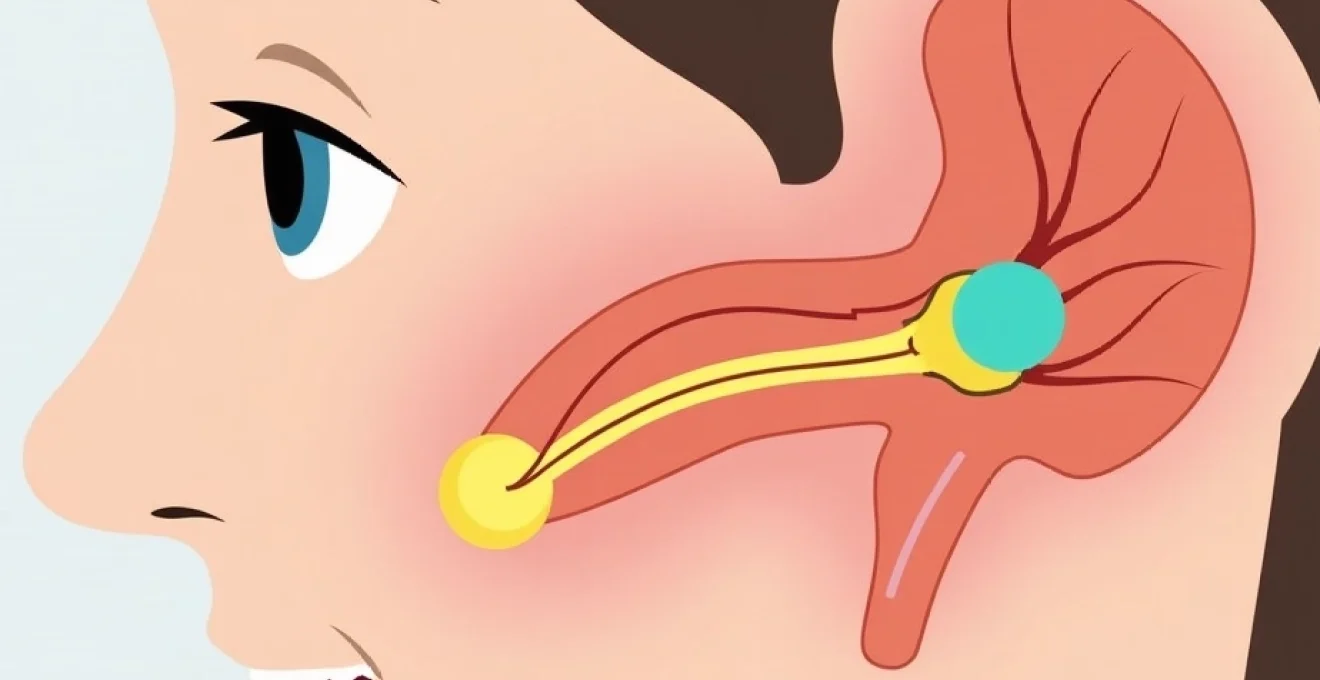
Adult tonsillectomy recovery presents unique challenges that differ significantly from paediatric procedures, with ear pain being one of the most distressing complications patients experience. This referred pain phenomenon affects the majority of adult patients following tonsil removal surgery, often causing greater discomfort than the actual surgical site itself. Understanding the neurological pathways responsible for this referred otalgia and implementing evidence-based management strategies can dramatically improve your recovery experience and reduce post-operative suffering.
The complexity of adult tonsillectomy recovery stems from the intricate nerve connections between the throat and ear structures, creating a cascade of referred sensations that can persist for up to two weeks post-surgery. Unlike direct ear infections or trauma, this type of auricular discomfort requires specialised management approaches that address both the underlying inflammatory processes and the specific nerve pathways involved in pain transmission.
Understanding referred otalgia following adult tonsillectomy
Referred otalgia represents one of the most poorly understood yet commonly experienced complications following adult tonsillectomy procedures. This phenomenon occurs when pain originating from the surgical site in the throat is perceived as coming from the ear itself, creating a confusing and often intense discomfort that can significantly impact your recovery journey. The underlying mechanism involves complex neurological pathways that transmit pain signals from the tonsillar fossa to the auditory system, resulting in ear pain that feels remarkably similar to acute otitis media.
Research indicates that approximately 85% of adult patients experience some degree of referred ear pain following tonsillectomy, with intensity typically peaking between days three to five post-operatively. This timing correlates directly with the inflammatory response cascade triggered by surgical tissue damage and the subsequent healing process. Understanding this timeline can help you prepare mentally for the recovery period and implement appropriate pain management strategies before the discomfort reaches its peak intensity.
Glossopharyngeal nerve pathway and pain transmission mechanisms
The glossopharyngeal nerve, designated as cranial nerve IX, serves as the primary conduit for referred ear pain following tonsillectomy procedures. This complex nerve structure originates from the medulla oblongata and provides sensory innervation to both the tonsillar region and portions of the middle ear cavity. When surgical trauma disrupts the tonsillar tissues, inflammatory mediators activate pain receptors that transmit signals along the glossopharyngeal nerve pathway, creating the sensation of deep ear pain despite the absence of any actual ear pathology.
The anatomical architecture of the glossopharyngeal nerve includes multiple branching patterns that explain why ear pain can manifest in different locations and intensities. The tympanic branch, specifically known as Jacobson’s nerve, carries sensory information from the middle ear mucosa and can become hyperactive following tonsillar surgery. This hyperactivity results in spontaneous pain signals that your brain interprets as originating from within the ear canal or deeper structures, creating a phantom pain sensation that can be remarkably convincing and distressing.
Trigeminal nerve branch involvement in Post-Surgical ear discomfort
The trigeminal nerve system contributes significantly to post-tonsillectomy ear pain through its auriculotemporal branch, which provides sensory innervation to the external ear canal and surrounding tissues. Following surgical trauma to the tonsillar region, inflammatory cytokines can create cross-sensitisation between different nerve pathways, causing the trigeminal system to become hyperresponsive to normal stimuli. This cross-sensitisation phenomenon explains why some patients experience ear pain that extends beyond the typical referred otalgia pattern.
The mandibular division of the trigeminal nerve shares anatomical connections with the glossopharyngeal system through the otic ganglion, creating potential points of convergence where pain signals can be amplified or modified. When you experience ear pain following tonsillectomy, multiple nerve pathways may be simultaneously activated, resulting in complex pain patterns that can include sharp, shooting sensations, dull aching, or burning discomfort that may radiate to the temporomandibular joint region.
Inflammatory response timeline and ear pain correlation
The inflammatory cascade following adult tonsillectomy follows a predictable timeline that directly correlates with the onset and intensity of referred ear pain. During the initial 24-48 hours post-surgery, prostaglandin E2 and other inflammatory mediators accumulate at the surgical site, sensitising nerve endings and lowering the threshold for pain signal transmission. This early inflammatory phase typically coincides with the onset of referred otalgia, which may initially be mild but progressively intensifies as the inflammatory response peaks.
Between days three to seven post-operatively, the inflammatory response reaches maximum intensity as neutrophils and macrophages infiltrate the healing tissues. During this critical period, you may experience the most severe ear pain, often accompanied by increased sensitivity to jaw movement, swallowing, and even light touch around the ear area. The formation of granulation tissue and the beginning of epithelialisation typically occur during the second week, corresponding with gradual resolution of referred ear pain as inflammatory mediators are cleared from the surgical site.
Distinguishing between referred pain and otitis media complications
Differentiating between referred otalgia and actual ear infections represents a critical diagnostic challenge that can significantly impact your treatment approach. Referred ear pain following tonsillectomy typically presents without the classic signs of otitis media, such as hearing loss, ear drainage, or visible changes to the tympanic membrane. The pain often worsens with swallowing or jaw movement, directly correlating with activation of the surgical site, whereas true otitis media pain remains relatively constant regardless of throat activity.
True post-operative ear infections, while rare, can occur as secondary complications of tonsillectomy procedures. These genuine infections typically present with purulent drainage, hearing impairment, and fever beyond the expected post-surgical inflammatory response. If you experience these additional symptoms alongside ear pain, immediate medical evaluation is essential to rule out actual otitis media or more serious complications such as necrotising otitis externa.
Immediate Post-Operative analgesic protocols for otalgia management
Effective management of post-tonsillectomy ear pain requires a multimodal approach that addresses both the inflammatory component and the neuropathic elements of referred otalgia. The optimal analgesic protocol combines scheduled anti-inflammatory medications with targeted pain relief strategies, implemented immediately following surgery to prevent the establishment of chronic pain pathways. Research demonstrates that proactive pain management during the first 72 hours post-operatively significantly reduces overall recovery time and improves long-term outcomes.
The concept of pre-emptive analgesia plays a crucial role in managing post-tonsillectomy ear pain, as early intervention can prevent the sensitisation of pain pathways that contribute to prolonged recovery periods. Modern pain management protocols emphasise the importance of maintaining consistent therapeutic drug levels rather than relying on as-needed dosing, which often allows pain to escalate beyond manageable levels before intervention occurs.
Paracetamol and ibuprofen combination therapy dosage guidelines
The cornerstone of post-tonsillectomy ear pain management involves the strategic combination of paracetamol and ibuprofen, which provides both analgesic and anti-inflammatory effects through complementary mechanisms of action. Paracetamol should be administered at 1000mg every six hours for adults, maintaining steady plasma levels that provide consistent pain relief without the gastrointestinal side effects associated with higher doses. This regular dosing schedule proves particularly effective for managing the constant, dull aching component of referred ear pain.
Ibuprofen administration at 400mg every eight hours provides targeted anti-inflammatory effects that address the root cause of referred otalgia while offering additional analgesic properties. The timing of ibuprofen doses should be staggered with paracetamol administration to maintain continuous pain coverage throughout the day. However, you should monitor for potential side effects including gastrointestinal irritation and ensure adequate hydration to support renal function during the recovery period.
Opioid-based pain relief: codeine and tramadol administration
For severe post-tonsillectomy ear pain that proves refractory to standard anti-inflammatory protocols, opioid medications may provide necessary relief during the acute recovery phase. Codeine phosphate, typically prescribed at 30-60mg every four to six hours, offers moderate analgesic effects while minimising the risk of respiratory depression associated with stronger opioids. The metabolism of codeine to morphine varies significantly between individuals, with approximately 10% of the population being poor metabolisers who may experience reduced efficacy.
Tramadol presents an alternative opioid option with additional noradrenergic and serotonergic properties that may prove particularly effective for neuropathic components of referred ear pain. The typical tramadol dosage ranges from 50-100mg every six hours, with a maximum daily dose of 400mg to prevent seizure risk. You should be aware that tramadol can interact with numerous medications and may cause serotonin syndrome when combined with certain antidepressants or other serotonergic drugs.
Topical anaesthetic applications: lidocaine gel and benzocaine solutions
Topical anaesthetic applications offer localised pain relief for post-tonsillectomy ear pain without the systemic effects associated with oral medications. Lidocaine gel applied directly to the external ear canal can provide temporary relief from superficial ear pain, particularly when the discomfort involves the auricular region. The application should be limited to small amounts to prevent systemic absorption, with effects typically lasting 30-60 minutes per application.
Benzocaine-containing ear drops may provide additional relief for deeper ear pain sensations, though their effectiveness for referred otalgia varies considerably between patients. These topical agents work by blocking sodium channels in nerve membranes, preventing the transmission of pain signals from peripheral receptors. You should avoid overuse of topical anaesthetics, as prolonged application can lead to contact sensitisation or masking of important warning signs that might indicate complications.
Corticosteroid use: prednisolone timing and Anti-Inflammatory effects
Corticosteroid therapy represents a powerful intervention for managing severe post-tonsillectomy inflammation and associated referred ear pain. Prednisolone, typically prescribed as a short course beginning on post-operative day three or four, can significantly reduce inflammatory mediator production and accelerate tissue healing. The standard regimen involves 20-30mg daily for three to five days, followed by a rapid taper to minimise side effects while maintaining therapeutic benefits.
The timing of corticosteroid initiation proves critical for optimal effectiveness, as early administration may interfere with normal healing processes while delayed treatment offers reduced anti-inflammatory benefits. Research suggests that corticosteroid therapy initiated during the peak inflammatory phase provides maximum pain relief and may reduce overall recovery time by 20-30%. However, you should be monitored for potential side effects including blood glucose elevation, mood changes, and delayed wound healing, particularly if you have underlying health conditions.
Cryotherapy and heat application techniques for auricular pain relief
Temperature modulation represents a non-pharmacological approach to managing post-tonsillectomy ear pain that can provide significant relief when implemented correctly. Cryotherapy applications work by reducing local inflammation and slowing nerve conduction velocity, effectively diminishing pain signal transmission from affected areas. The application of cold therapy should be limited to 15-20 minute intervals to prevent tissue damage while maximising analgesic benefits through the gate control theory of pain modulation.
Ice pack applications to the external ear and surrounding tissues can provide immediate relief from acute ear pain episodes, particularly during the first few days following surgery when inflammatory responses peak. The cold temperature causes vasoconstriction, reducing inflammatory mediator circulation and providing temporary numbing effects that can break the cycle of pain and muscle tension. You should wrap ice packs in thin towels to prevent direct skin contact and frostbite injury, while maintaining consistent application pressure to ensure effective heat transfer.
Heat therapy offers complementary benefits to cryotherapy by promoting vasodilation and muscle relaxation, which can be particularly effective for managing deeper, aching ear pain that characterises the later stages of recovery. Warm compress applications at temperatures between 40-45°C provide optimal therapeutic benefits without risk of thermal injury. The heat promotes increased blood flow to the affected area, facilitating the removal of inflammatory metabolites while delivering nutrients necessary for tissue repair. Alternating between cold and heat therapy in 15-minute cycles can provide enhanced pain relief compared to single-modality temperature therapy.
The effectiveness of temperature therapy can be enhanced through proper positioning and timing of applications. Lying on the unaffected side while applying temperature therapy to the painful ear allows gravity to assist with circulation and reduces pressure on sensitive tissues. You should coordinate temperature therapy applications with medication schedules to provide comprehensive pain coverage during peak discomfort periods, typically in the morning hours when referred ear pain tends to be most severe.
Hydration strategies and electrolyte balance for accelerated healing
Maintaining optimal hydration status represents a fundamental component of post-tonsillectomy recovery that directly impacts both healing rates and pain management outcomes. Adequate fluid intake supports the body’s natural inflammatory response resolution while preventing the complications associated with dehydration, such as increased pain sensitivity and delayed tissue repair. Adult patients require a minimum of 2.5-3 litres of fluid daily during the acute recovery phase, with additional requirements based on individual factors such as body weight, ambient temperature, and activity level.
The relationship between hydration and referred ear pain involves multiple physiological mechanisms, including maintenance of optimal blood viscosity for inflammatory mediator clearance and prevention of secondary complications that can exacerbate pain perception. Dehydration can lead to increased plasma osmolality, which affects nerve membrane stability and potentially increases pain signal transmission. Additionally, adequate hydration supports kidney function, ensuring efficient clearance of pain medications and their metabolites to prevent drug accumulation and associated side effects.
Isotonic solution benefits: normal saline and hartmann’s solution
Isotonic solutions provide optimal electrolyte replacement for patients experiencing significant fluid losses due to reduced oral intake, fever, or medication-induced diuresis. Normal saline (0.9% sodium chloride) offers immediate rehydration benefits while maintaining plasma osmolality within physiological ranges. However, excessive normal saline administration can lead to hyperchloraemic acidosis, making Hartmann’s solution a preferred alternative for patients requiring substantial fluid replacement during recovery.
Hartmann’s solution provides a more physiologically balanced electrolyte profile, containing sodium, chloride, potassium, and lactate in concentrations that closely mirror plasma composition. This balanced formulation supports optimal cellular function while preventing the electrolyte imbalances that can contribute to increased pain sensitivity and delayed healing. For patients experiencing severe dehydration or inability to maintain oral intake, intravenous isotonic solutions may be necessary to restore fluid balance and support recovery processes.
Anti-dehydration protocols: ORS and IV fluid requirements
Oral rehydration solutions (ORS) represent the first-line treatment for mild to moderate dehydration in post-tonsillectomy patients who can tolerate oral intake. Commercial ORS formulations contain precisely balanced glucose and electrolytes that optimise intestinal fluid absorption through sodium-glucose co-transport mechanisms. The standard WHO/UNICEF ORS formulation provides 75mEq/L sodium, 65mEq/L chloride, 20mEq/L potassium, and 75mmol/L glucose, creating an osmolality of 245mOsm/L that maximises absorption efficiency.
Intravenous fluid requirements for post-tonsillectomy patients depend on multiple factors including baseline hydration status, ongoing losses, and comorbid conditions. The standard maintenance fluid requirement of 25-30ml/kg/day should be increased by 10-15% during the acute inflammatory phase to account for increased metabolic demands and potential losses through fever or reduced oral intake. Monitoring urine output, skin turgor, and mucous membrane moisture provides reliable indicators of hydration status and guides fluid replacement strategies.
Throat moistening techniques to reduce referred ear pressure
Maintaining optimal throat moisture levels can significantly reduce the intensity of referred ear pain by preventing secondary inflammation and irritation of already sensitised nerve pathways. Humidification of inspired air through steam inhalation or mechanical humidifiers maintains mucosal moisture and prevents the desiccation that can exacerbate post-surgical inflammation. Target humidity levels between 40-60% provide optimal comfort without promoting bacterial growth or condensation issues.
Throat irrigation with warm saline solutions offers direct moistening benefits while providing mild analgesic effects through osmotic and temperature mechanisms. A solution of 0.9% sodium chloride warmed to body temperature can be used for gentle gargling or throat irrigation every 2-3 hours during waking periods. The mechanical action of gentle gargling promotes circulation to the healing tissues while removing debris and inflammatory exudates that can contribute to pain and delayed healing.
Sleep positioning and environmental modifications for comfort optimisation
Sleep quality significantly impacts pain perception and healing rates following tonsillectomy,
making proper sleep positioning critical for managing referred ear pain and promoting healing. Elevating your head 30-45 degrees above heart level reduces venous congestion and inflammatory fluid accumulation around the surgical site, which can significantly diminish the intensity of referred otalgia. This positioning utilises gravity to promote lymphatic drainage while reducing pressure on sensitised nerve pathways that contribute to ear pain transmission.
Side-sleeping positions require careful consideration during tonsillectomy recovery, as lying on the affected side can exacerbate ear pain through direct pressure and positioning-induced nerve compression. Sleeping on the unaffected side allows gravity to assist with drainage of inflammatory exudates while reducing mechanical stress on healing tissues. However, alternating sides periodically prevents the development of pressure sores and maintains optimal circulation to both ears throughout the night.
Environmental modifications play an equally important role in optimising sleep quality and pain management. Maintaining bedroom humidity between 40-60% prevents mucosal desiccation that can worsen throat pain and subsequently increase referred ear discomfort. White noise machines or soft background sounds can help mask the tinnitus-like sensations that sometimes accompany referred otalgia, promoting deeper sleep phases that are essential for tissue repair and pain modulation.
Temperature regulation within the sleep environment affects both comfort and healing rates, with optimal bedroom temperatures ranging between 18-21°C. Cooler environments support the natural circadian decrease in core body temperature that promotes sleep onset, while preventing overheating that can exacerbate post-surgical inflammation. Using breathable bedding materials and moisture-wicking pillowcases can further enhance comfort by managing perspiration and maintaining optimal skin temperature around the head and neck region.
Red flag symptoms requiring immediate ENT specialist consultation
Recognising potentially serious complications that can masquerade as routine post-tonsillectomy ear pain represents a critical aspect of safe recovery management. While referred otalgia is expected following tonsil removal, certain warning signs indicate complications requiring immediate medical intervention to prevent serious sequelae. Understanding these red flag symptoms empowers you to seek appropriate care before minor complications progress to life-threatening conditions.
Post-tonsillectomy haemorrhage represents the most serious complication, occurring in approximately 2-5% of adult patients and potentially manifesting as severe ear pain accompanied by bleeding. Primary haemorrhage occurs within the first 24 hours post-operatively, while secondary bleeding typically develops 5-10 days after surgery when eschar separation occurs. Any visible blood in saliva, persistent metallic taste, or sudden onset of severe ear pain accompanied by throat discomfort warrants immediate emergency department evaluation.
Severe, unilateral ear pain that develops suddenly and proves refractory to prescribed pain medications may indicate necrotising otitis externa or other serious infectious complications. This condition, while rare, can progress rapidly in immunocompromised patients or those with diabetes, potentially causing permanent hearing loss or intracranial extension. The pain typically exhibits a boring, deep quality that worsens progressively and may be accompanied by purulent ear drainage or facial nerve weakness.
Fever patterns provide crucial diagnostic information for distinguishing between normal post-operative responses and serious complications. Temperature elevations above 38.5°C (101.3°F) persisting beyond 48 hours post-operatively, or any fever developing after an initial afebrile period, suggest possible infection requiring antibiotic intervention. Rigors, night sweats, or systemic symptoms accompanying fever indicate potential bacteraemia or abscess formation that demands immediate medical evaluation.
Neurological symptoms accompanying ear pain represent particularly concerning developments that may indicate intracranial complications or cranial nerve involvement. Facial weakness, visual disturbances, severe headache, neck stiffness, or altered mental status occurring in conjunction with ear pain require emergency neurological assessment. These symptoms may herald conditions such as meningitis, brain abscess, or cavernous sinus thrombosis that can develop as rare but serious complications of upper respiratory tract surgery.
Sudden hearing loss or significant hearing impairment developing after tonsillectomy represents another red flag requiring urgent ENT evaluation. While mild conductive hearing loss may occur due to eustachian tube inflammation, sudden sensorineural hearing loss indicates potential inner ear complications that require immediate intervention to preserve hearing function. Accompanying symptoms such as vertigo, tinnitus, or balance disturbances further emphasise the urgency of specialist consultation.
Respiratory distress or airway compromise can manifest initially as severe ear and throat pain before progressing to life-threatening obstruction. Tongue swelling, difficulty swallowing secretions, or progressive voice changes may herald laryngeal oedema or other airway complications requiring immediate intervention. The development of stridor, increased respiratory rate, or accessory muscle use demands immediate emergency medical attention regardless of other symptom severity.
Dehydration symptoms often accompany severe pain that prevents adequate oral intake, potentially leading to serious metabolic complications. Signs including decreased urine output, dark urine colour, dry mucous membranes, skin tenting, or orthostatic symptoms indicate significant fluid loss requiring medical evaluation. Severe dehydration can impair wound healing, increase infection risk, and exacerbate pain perception through multiple physiological mechanisms.
Understanding the timeline of normal versus concerning symptoms helps differentiate between expected recovery patterns and complications requiring intervention. Pain that continues to worsen beyond post-operative day 5-7, or ear pain that develops for the first time more than one week after surgery, suggests potential complications rather than normal referred otalgia. Similarly, any new symptom development after initial improvement warrants medical evaluation to rule out secondary complications or treatment failures.