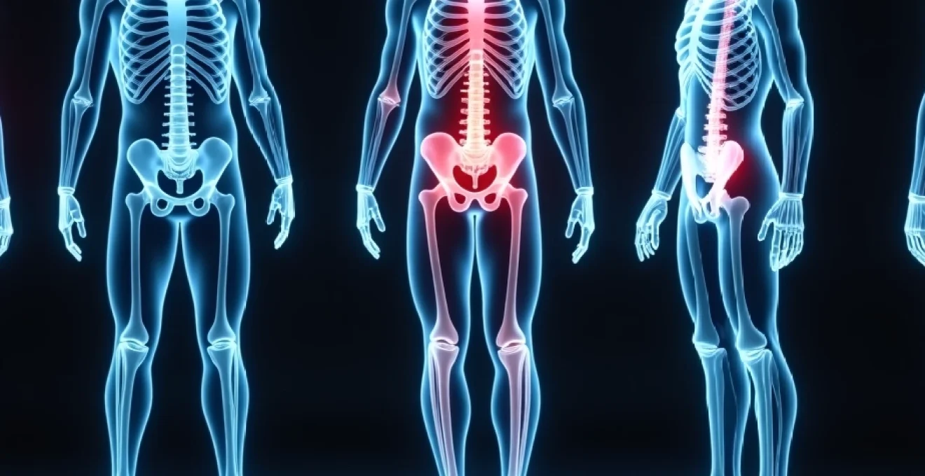
The relationship between medical imaging and soft tissue visualisation remains one of the most frequently misunderstood aspects of diagnostic medicine. When patients experience joint injuries, particularly those involving potential ligament damage, understanding the capabilities and limitations of different imaging modalities becomes crucial for appropriate treatment decisions. X-ray imaging, whilst exceptional for bone assessment, faces significant technical challenges when attempting to visualise ligamentous structures, fundamentally altering the diagnostic approach for soft tissue injuries.
X-ray imaging limitations in soft tissue visualisation
X-ray technology operates on fundamental principles of radiation attenuation that create inherent limitations for soft tissue assessment. The electromagnetic radiation used in conventional radiography interacts differently with various tissue types based on their atomic composition and density, making ligaments virtually invisible on standard X-ray images.
Radiodensity properties of ligamentous structures
Ligaments possess remarkably low radiodensity compared to osseous structures, rendering them essentially transparent to conventional X-ray beams. These fibrous connective tissues contain predominantly collagen fibres, water, and proteoglycans, none of which significantly attenuate X-ray photons. The atomic number of elements within ligamentous tissue remains consistently low, primarily comprising carbon, hydrogen, oxygen, and nitrogen, which interact minimally with diagnostic X-ray energies typically ranging from 50-120 kilovolts peak.
Research indicates that ligaments demonstrate radiographic opacity values similar to surrounding soft tissues, creating an indistinguishable grey appearance on conventional radiographs. This similarity in attenuation coefficients means that even healthy, intact ligaments cannot be differentiated from adjacent muscle, fat, or other connective tissues using standard radiographic techniques.
Contrast resolution challenges in conventional radiography
The contrast resolution limitations of X-ray imaging present significant obstacles for ligament visualisation. Contrast resolution refers to the ability to distinguish between tissues with similar radiographic densities, a critical factor for soft tissue assessment. Conventional radiography typically achieves contrast resolutions of approximately 1-2%, insufficient for distinguishing subtle differences between ligamentous structures and surrounding tissues.
Digital radiography systems have improved contrast resolution compared to film-screen combinations, yet they still cannot overcome the fundamental physics limitations inherent in X-ray imaging of soft tissues. Even with advanced post-processing algorithms and enhanced digital manipulation, ligaments remain invisible due to their inherently low contrast characteristics.
Photoelectric absorption coefficients in connective tissues
The photoelectric absorption coefficient for ligamentous tissue remains approximately 100 times lower than cortical bone at diagnostic X-ray energies. This dramatic difference explains why bones appear distinctly white on radiographs whilst soft tissues, including ligaments, appear as various shades of grey with minimal differentiation between structures.
Photoelectric interactions depend heavily on the atomic number of absorbing materials raised to the third or fourth power. Since ligaments contain primarily low atomic number elements, they demonstrate minimal photoelectric absorption, contributing to their radiographic transparency. This physical limitation cannot be overcome through technical adjustments or equipment modifications.
Beam attenuation differences between bone and soft tissue
Quantitative analysis reveals that cortical bone attenuates X-ray beams approximately 4-5 times more effectively than soft tissues at standard diagnostic energies. Ligaments, being composed primarily of collagen and water, demonstrate attenuation coefficients nearly identical to muscle tissue, creating virtually no discernible contrast on conventional radiographs.
Beam hardening effects further complicate soft tissue visualisation, as polychromatic X-ray beams become progressively harder as they traverse tissue. This phenomenon reduces the already minimal contrast differences between ligamentous structures and surrounding soft tissues, making reliable ligament assessment impossible through conventional radiographic means.
Advanced imaging modalities for ligament assessment
Modern medical imaging has evolved beyond conventional radiography to provide sophisticated alternatives for ligament evaluation. These advanced modalities utilise different physical principles to overcome the limitations inherent in X-ray imaging, enabling detailed assessment of soft tissue structures previously invisible to conventional radiographic techniques.
Magnetic resonance imaging protocols for ACL and PCL evaluation
Magnetic resonance imaging represents the gold standard for ligament assessment, particularly for crucial knee stabilisers such as the anterior and posterior cruciate ligaments. MRI protocols specifically designed for knee ligament evaluation typically employ multiple imaging sequences to optimise soft tissue contrast and spatial resolution.
Proton density-weighted sequences with fat saturation provide exceptional contrast between ligamentous structures and surrounding tissues, clearly delineating normal ligament architecture from pathological changes. T2-weighted sequences excel at identifying fluid accumulation and inflammatory changes associated with acute ligament injuries, whilst gradient echo sequences offer superior spatial resolution for detecting partial tears.
Three-dimensional MRI acquisitions enable multiplanar reconstructions aligned with specific ligament orientations, improving diagnostic accuracy for complex ligamentous injuries. Studies demonstrate that modern MRI protocols achieve sensitivities exceeding 95% for complete ACL tears and approximately 85% for partial injuries, representing substantial improvements over conventional imaging methods.
Ultrasonography applications in lateral collateral ligament diagnosis
High-resolution ultrasonography provides real-time assessment of superficial ligaments, particularly advantageous for evaluating lateral collateral ligament integrity in various joints. Modern ultrasound systems operating at frequencies between 10-15 MHz achieve spatial resolutions approaching 0.1 millimetres, sufficient for detailed ligament morphology assessment.
Dynamic ultrasound examination offers unique advantages for functional ligament assessment, enabling real-time visualisation during stress testing procedures. This capability proves particularly valuable for detecting subtle instability patterns that might remain undetected on static imaging studies. Colour Doppler imaging adds another dimension by identifying hypervascularity associated with acute ligament injuries and inflammatory responses.
Ultrasound-guided interventions, including therapeutic injections and needle placement procedures, benefit from real-time visualisation capabilities unavailable with other imaging modalities. The portability and cost-effectiveness of ultrasound systems make them increasingly attractive for immediate injury assessment in clinical and sporting environments.
CT arthrography techniques for glenohumeral joint ligaments
Computed tomography arthrography combines the cross-sectional imaging capabilities of CT with intra-articular contrast enhancement to improve ligament visualisation. This technique proves particularly effective for assessing glenohumeral joint capsular ligaments and labral structures that remain challenging to evaluate with conventional imaging methods.
Multidetector CT systems enable rapid acquisition of high-resolution datasets with isotropic voxel dimensions, facilitating multiplanar reconstructions in any desired orientation. The introduction of iodinated contrast material into joint spaces dramatically improves soft tissue contrast, allowing detailed assessment of capsular ligaments and associated structures.
Three-dimensional volume rendering techniques provide surgeons with invaluable preoperative planning information, particularly for complex shoulder reconstruction procedures. CT arthrography demonstrates superior sensitivity for detecting labral tears and capsular ligament injuries compared to conventional MRI in certain clinical scenarios.
MR arthrography advantages in SLAP lesion detection
Magnetic resonance arthrography represents the pinnacle of soft tissue imaging for certain applications, combining the superior soft tissue contrast of MRI with the enhanced detail provided by intra-articular contrast administration. This technique proves particularly valuable for detecting superior labrum anterior-posterior (SLAP) lesions and associated biceps anchor pathology.
Gadolinium-based contrast agents provide T1 shortening effects that dramatically improve the conspicuity of labral and ligamentous structures on appropriately weighted sequences. The distension effect of contrast injection helps separate closely approximated structures, improving detection of subtle tears and degenerative changes that might remain occult on conventional MRI studies.
Recent advances in MR arthrography include the development of specialised coil arrays and pulse sequences optimised for small joint imaging. These innovations have expanded the technique’s applications to include wrist, ankle, and other peripheral joint assessments where detailed ligament evaluation proves clinically essential.
Indirect x-ray signs of ligamentous pathology
Although X-rays cannot directly visualise ligaments, experienced radiologists and clinicians recognise numerous indirect radiographic signs that suggest ligamentous injury or instability. These secondary findings often provide valuable diagnostic information when interpreted within appropriate clinical contexts, enabling informed treatment decisions despite the inherent limitations of conventional radiography.
Joint space widening in scapholunate ligament injuries
Scapholunate ligament disruption frequently manifests as pathological widening of the scapholunate interval on posteroanterior wrist radiographs. Normal scapholunate distance measures approximately 2-3 millimetres, whilst measurements exceeding 5 millimetres strongly suggest significant ligamentous injury. This radiographic finding, often termed the “Terry Thomas sign,” represents one of the most reliable indirect indicators of wrist ligament pathology visible on conventional X-rays.
Dynamic stress radiographs enhance the sensitivity of X-ray examination for detecting scapholunate ligament injuries by applying controlled forces that exacerbate underlying instability. Clenched-fist radiographs create physiological loading conditions that may unmask subtle ligamentous injuries not apparent on standard static images.
Sequential radiographic measurements during healing provide objective documentation of treatment progress and guide decisions regarding return to activity. Research demonstrates that restoration of normal scapholunate intervals correlates strongly with functional outcomes following ligament reconstruction procedures.
Stress radiography protocols for ankle syndesmotic assessment
Ankle syndesmotic ligament injuries, commonly occurring in rotational ankle injuries, require specialised radiographic techniques for accurate assessment. Stress radiography involves applying controlled external forces during X-ray exposure to demonstrate pathological joint separation that might remain undetected on standard radiographs.
External rotation stress tests specifically target the anterior tibiofibular ligament and interosseous membrane, creating measurable separation between the tibia and fibula when these structures are compromised. Quantitative measurements of tibiofibular clear space, tibiofibular overlap, and medial clear space provide objective criteria for diagnosing syndesmotic injuries using conventional radiographic techniques.
Bilateral comparison radiographs prove essential for accurate interpretation, as normal anatomical variations can simulate pathological findings. The contralateral uninjured ankle serves as an internal control, improving diagnostic accuracy and reducing false-positive interpretations of normal anatomical variants.
Avulsion fracture patterns at ligament insertion sites
Ligament injuries occasionally result in avulsion fractures at bony attachment sites, creating distinctive radiographic patterns that suggest the nature and severity of underlying soft tissue damage. These bony fragments represent the only direct radiographic evidence of ligamentous trauma visible on conventional X-rays, making their recognition crucial for accurate diagnosis.
Pellegrini-Stieda lesions demonstrate heterotopic ossification following medial collateral ligament injuries in the knee, appearing as characteristic calcifications along the medial femoral condyle on radiographs obtained weeks to months following injury. Similarly, anterior cruciate ligament injuries may produce avulsion fractures of the tibial eminence, particularly in younger patients with immature skeletons.
The recognition of characteristic avulsion patterns enables clinicians to infer the presence of significant ligamentous injury even when direct soft tissue visualisation remains impossible through conventional radiographic means.
Bone displacement measurements in acromioclavicular joint injuries
Acromioclavicular joint injuries demonstrate characteristic displacement patterns on conventional radiographs that correlate with the degree of ligamentous damage. Measurement of coracoclavicular distance provides quantitative assessment of injury severity, with increases exceeding 25-50% compared to the contralateral side indicating significant ligament disruption.
Weight-bearing or stress radiographs enhance the sensitivity of X-ray examination for detecting acromioclavicular ligament injuries by accentuating displacement patterns that might remain subtle on standard images. The application of downward traction or weights during radiographic examination helps differentiate between partial and complete ligament ruptures based on the degree of observed displacement.
Serial radiographic monitoring enables objective assessment of treatment effectiveness and guides decisions regarding surgical intervention. Progressive displacement measurements provide valuable prognostic information and help determine the optimal timing for therapeutic interventions.
Clinical applications and diagnostic workflow
The integration of X-ray imaging within comprehensive diagnostic algorithms for suspected ligament injuries requires careful consideration of clinical presentation, mechanism of injury, and examination findings. Understanding when conventional radiography provides valuable information versus when advanced imaging becomes necessary represents a crucial clinical skill that directly impacts patient outcomes and healthcare resource utilisation.
Emergency department protocols typically initiate evaluation with conventional radiographs to exclude fractures and obvious joint dislocations before proceeding to more sophisticated imaging studies. This approach proves particularly important in acute trauma settings where bony injuries require immediate recognition and management. However, clinicians must recognise that negative X-ray findings do not exclude significant ligamentous injuries, necessitating clinical correlation and often requiring additional imaging studies.
Cost-effectiveness considerations influence diagnostic decision-making, as conventional radiographs cost significantly less than advanced imaging modalities whilst providing essential information about bony structures. Studies indicate that X-ray examination costs approximately one-tenth the price of MRI studies, making initial radiographic screening an economically sound approach when guided by appropriate clinical algorithms.
The temporal aspects of ligament injury evolution affect imaging strategy selection, as certain radiographic signs develop gradually over time whilst others manifest immediately following injury. Understanding these temporal relationships enables clinicians to optimise imaging timing and modality selection for maximum diagnostic yield whilst minimising patient radiation exposure and healthcare costs.
Radiographic contrast agents and ligament visualisation techniques
Specialised radiographic techniques utilising contrast enhancement represent attempts to overcome the inherent limitations of conventional X-ray imaging for soft tissue assessment. These methods, whilst not widely employed in routine clinical practice, demonstrate the ongoing evolution of radiographic technology and its potential applications for ligament evaluation in specific clinical scenarios.
Arthrography involves the injection of contrast material directly into joint spaces to outline synovial structures and capsular ligaments. Double-contrast arthrography combines positive contrast agents with air or carbon dioxide to create optimal visualisation conditions for certain ligamentous structures. This technique proved particularly valuable before the widespread availability of MRI and continues to serve specific clinical applications where real-time fluoroscopic guidance provides advantages over static imaging methods.
Tenography and bursography represent related contrast-enhanced techniques that may indirectly assess ligamentous structures through their relationships with adjacent synovial spaces and tendon sheaths. These procedures require specialised expertise and carry inherent risks associated with invasive contrast injection, limiting their routine clinical application to cases where non-invasive alternatives prove inadequate.
Digital subtraction techniques attempt to enhance soft tissue contrast by electronically removing bony structures from radiographic images, theoretically improving ligament visualisation. However, practical limitations including motion artefacts and incomplete bone subtraction have prevented widespread adoption of these methods for routine ligament assessment.
The development of novel contrast agents specifically designed for soft tissue enhancement represents an active area of radiographic research. Nanoparticle-based contrast materials show promise for improving ligament visualisation through targeted accumulation in connective tissues, though clinical applications remain largely experimental at this time.
Future developments in radiographic technology may eventually overcome current limitations through advanced detector systems, novel contrast agents, and sophisticated image processing algorithms specifically designed for soft tissue enhancement.