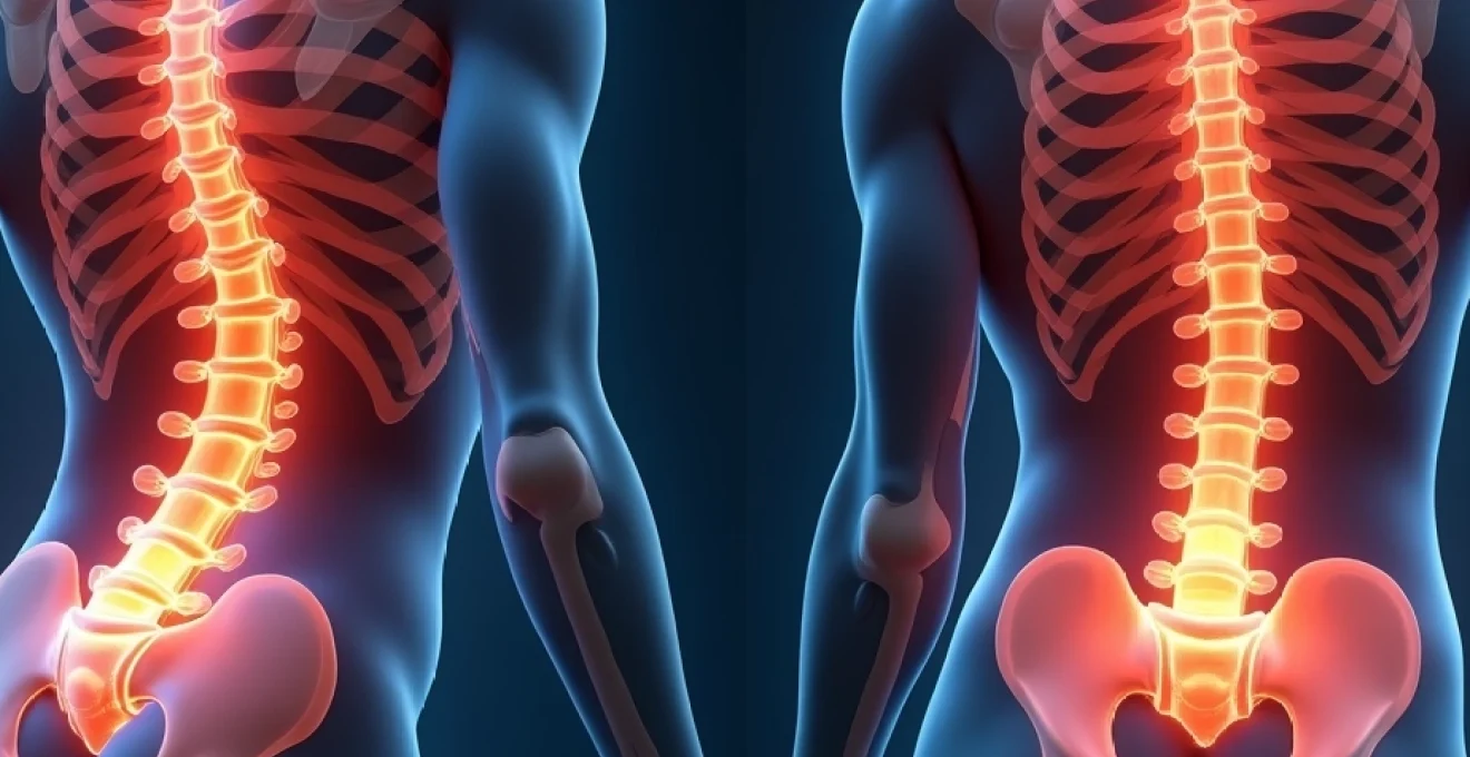
The lumbosacral junction represents one of the most biomechanically demanding regions of the human spine, where the fifth lumbar vertebra meets the first sacral segment. An annular fissure at this critical juncture involves a tear in the tough outer ring of the intervertebral disc, known as the annulus fibrosus. This condition affects millions of people worldwide and can significantly impact quality of life through persistent pain and functional limitations. Understanding the complex pathophysiology, clinical presentation, and treatment options for L5-S1 annular fissures is essential for both healthcare providers and patients navigating this challenging condition.
Understanding annular fissure pathophysiology at the lumbosacral junction
Annulus fibrosus structural composition and collagen architecture
The annulus fibrosus functions as the primary structural component responsible for containing the nucleus pulposus and distributing mechanical loads across the disc space. This remarkable structure consists of 15 to 20 concentric lamellae, each composed primarily of type I collagen fibres arranged in alternating oblique patterns. The unique criss-cross architecture provides exceptional tensile strength whilst allowing controlled movement between vertebral segments.
The collagen fibres in each lamella are oriented approximately 30 degrees from the horizontal plane, with adjacent layers running in opposite directions. This sophisticated arrangement creates a natural shock-absorbing system capable of withstanding significant compressive, tensile, and torsional forces. However, the posterolateral region of the annulus fibrosus demonstrates inherent weakness due to thinner lamellae and reduced collagen density, making it particularly susceptible to tear formation.
Degenerative cascade mechanisms in L5-S1 disc degeneration
Disc degeneration at the L5-S1 level follows a predictable cascade of biochemical and structural changes. The process typically begins with reduced proteoglycan content in the nucleus pulposus, leading to decreased water retention and subsequent loss of disc height. As hydration diminishes, the nucleus becomes increasingly fibrous and less capable of distributing loads evenly across the annular structure.
Research indicates that matrix metalloproteinases play a crucial role in breaking down collagen and proteoglycans within the disc matrix. Simultaneously, inflammatory cytokines such as interleukin-1β and tumour necrosis factor-α accelerate the degenerative process. This inflammatory environment not only weakens the structural integrity of the annulus fibrosus but also sensitises nerve endings, contributing to the characteristic pain associated with annular fissures.
Biomechanical stress distribution at the lumbosacral transition
The L5-S1 segment experiences unique biomechanical stresses due to its position at the junction between the mobile lumbar spine and the rigid sacrum. This transitional zone must accommodate significant shear forces during normal activities, with intradiscal pressures reaching up to 2.3 MPa during heavy lifting. The lumbosacral angle, typically measuring 140 degrees, creates additional anterior shear stress that can predispose the posterior annulus to failure.
Studies demonstrate that repetitive loading cycles, rather than single traumatic events, are responsible for most annular fissures. The phenomenon of fatigue failure occurs when accumulated microdamage exceeds the tissue’s repair capacity. This explains why annular tears often develop insidiously in individuals with occupations requiring frequent bending, lifting, or prolonged sitting positions.
Inflammatory mediator release and nociceptive pathway activation
When an annular fissure develops, the release of inflammatory mediators triggers a complex cascade of nociceptive responses. The outer third of the annulus fibrosus contains numerous nerve endings supplied by the sinuvertebral nerve, making these tears particularly painful when they extend to this region. Substance P and other neuropeptides are released from damaged nerve terminals, amplifying the inflammatory response and creating a cycle of pain and tissue damage.
The inflammatory process also increases vascular permeability, allowing immune cells to infiltrate the normally avascular disc space. This immune response, whilst intended to clear damaged tissue, can perpetuate inflammation and delay healing. Understanding this pathophysiology has led to targeted treatments aimed at interrupting the inflammatory cascade and promoting tissue repair.
Clinical manifestation patterns and diagnostic criteria for L5-S1 annular tears
Discogenic pain presentation and provocative testing protocols
Discogenic pain from L5-S1 annular fissures presents with distinctive characteristics that help differentiate it from other causes of lower back pain. Patients typically report deep, aching pain localised to the lower lumbar region with occasional radiation to the buttocks or posterior thigh. The pain often worsens with prolonged sitting, forward flexion, or coughing and sneezing, activities that increase intradiscal pressure.
Provocative testing protocols play a crucial role in identifying discogenic pain sources. The discogenic pain provocation test involves sustained forward flexion for 60 seconds, which typically reproduces or exacerbates symptoms in patients with annular fissures. Additionally, the centralization phenomenon, where peripheral pain moves towards the midline with repeated extension exercises, can help distinguish discogenic pain from radicular symptoms.
Neurological deficit assessment in S1 radiculopathy
When annular fissures extend sufficiently to compress the S1 nerve root, patients may develop characteristic neurological deficits. S1 radiculopathy typically presents with weakness in plantarflexion and eversion of the foot, diminished or absent Achilles reflex, and sensory disturbances along the lateral foot and little toe. These findings can be subtle in early stages but become more pronounced as nerve compression progresses.
Assessment of S1 nerve root function should include manual muscle testing of the gastrocnemius and soleus muscles, evaluation of single heel-rise ability, and comprehensive sensory examination of the S1 dermatome. The straight leg raise test, whilst not specific for annular fissures, may reproduce radicular symptoms when nerve root irritation is present. A positive test typically occurs at less than 60 degrees of hip flexion and reproduces the patient’s familiar leg pain.
Differential diagnosis from piriformis syndrome and sacroiliac joint dysfunction
Distinguishing L5-S1 annular fissures from other common causes of lower back and leg pain requires careful clinical assessment. Piriformis syndrome can mimic S1 radiculopathy but typically responds to specific stretching manoeuvres and lacks the centralization pattern seen with discogenic pain. The FAIR test (flexion, adduction, and internal rotation) often reproduces symptoms in piriformis syndrome but remains negative in pure discogenic pain.
Sacroiliac joint dysfunction presents another diagnostic challenge, as it can coexist with disc pathology. Pain from sacroiliac joint dysfunction typically localises more laterally and may respond to specific joint mobilisation techniques. Provocative tests such as the FABER test and posterior shear test can help identify sacroiliac joint involvement, though these conditions frequently occur together in complex pain syndromes.
Pain distribution mapping and dermatomal analysis
Accurate pain mapping provides valuable diagnostic information for L5-S1 annular fissures. Discogenic pain typically follows a non-dermatomal distribution pattern, often described as deep and difficult to localise precisely. Patients may use descriptors such as “burning,” “aching,” or “stabbing” to characterise their symptoms. The pain often varies in intensity throughout the day and may be influenced by posture and activity levels.
When radicular involvement occurs, pain distribution follows the S1 dermatome more precisely, extending from the posterior buttock down the posterior and lateral thigh to the lateral leg and foot. However, referred pain patterns can complicate diagnosis, as visceral structures and other spinal segments can produce similar pain distributions. Detailed pain mapping, including timing, triggers, and relieving factors, helps clinicians distinguish between different pain generators.
Advanced imaging modalities for annular fissure identification
High-intensity zone detection on T2-Weighted MRI sequences
Magnetic resonance imaging remains the gold standard for visualising annular fissures, with T2-weighted sequences providing optimal contrast for detecting high-intensity zones (HIZ) within the posterior annulus fibrosus. These HIZ appear as bright signal areas against the dark background of the normal annulus and represent areas of tissue disruption with increased water content. Research indicates that HIZ detection correlates with concordant pain reproduction during discography in approximately 87% of cases.
The classification of annular fissures on MRI follows specific criteria based on signal intensity and location. Grade 1 fissures show mild signal increase confined to the inner annular layers, whilst Grade 3 fissures extend through the full thickness of the annulus to the outer edge. T2-weighted fat-suppressed sequences enhance the conspicuity of small fissures and help differentiate them from normal anatomical variants.
CT discography protocols and concordant pain reproduction
Computed tomography discography represents the most specific diagnostic test for identifying painful annular fissures. This procedure involves injection of contrast material directly into the disc space under fluoroscopic guidance, followed by CT imaging to visualise the internal disc architecture. The key diagnostic criterion is reproduction of the patient’s familiar pain pattern during injection, termed concordant pain reproduction .
Modern discography protocols emphasise the importance of control level testing to ensure specificity. Adjacent normal-appearing discs are also injected, and a positive test requires that only the suspected symptomatic level reproduces familiar pain. Pressure monitoring during injection provides additional diagnostic information, with abnormally low pressures often indicating significant structural compromise of the annulus fibrosus.
Magnetic resonance neurography in nerve root compression assessment
Magnetic resonance neurography utilises specialised sequences to enhance visualisation of peripheral nerves and detect subtle signs of nerve root irritation or compression. This advanced imaging technique can identify nerve root oedema, fascicular disruption, and altered signal intensity that may not be apparent on conventional MRI sequences. For L5-S1 annular fissures with suspected radicular involvement, neurography sequences can provide crucial information about the degree and location of nerve compromise.
The technique employs diffusion-weighted imaging and T2-weighted sequences with fat suppression to highlight nerve tissue contrast. Quantitative measurements of nerve cross-sectional area and signal intensity can objectively assess the severity of nerve involvement. This information proves particularly valuable when planning treatment strategies and predicting surgical outcomes.
Ultrasound-guided diagnostic injection techniques
Ultrasound guidance has emerged as a valuable tool for diagnostic injections around the L5-S1 segment, offering real-time visualisation without radiation exposure. This technique proves particularly useful for targeted injections into the facet joints, sacroiliac joints, and epidural space to help differentiate pain sources. The ability to visualise needle placement in real-time enhances safety and accuracy of diagnostic procedures.
Recent advances in ultrasound technology have improved visualisation of deep spinal structures, though the technique remains limited for direct disc visualisation due to bony interference. However, ultrasound-guided epidural injections can provide valuable diagnostic and therapeutic information when evaluating patients with suspected radicular symptoms from annular fissures.
Conservative treatment protocols and interventional pain management
Conservative management represents the first-line approach for most patients with L5-S1 annular fissures, with evidence supporting a multimodal strategy combining pain control, movement restoration, and activity modification. The initial focus centres on reducing inflammation and controlling pain whilst protecting the injured tissue from further damage. Non-steroidal anti-inflammatory drugs (NSAIDs) provide both analgesic and anti-inflammatory effects, though their use requires careful consideration of gastrointestinal and cardiovascular risks, particularly in long-term treatment scenarios.
Physical therapy protocols specifically designed for discogenic pain emphasise postural correction, core stabilisation, and movement retraining. The McKenzie method has shown particular efficacy in managing discogenic pain through directional preference exercises that promote centralization of symptoms. Research demonstrates that patients who achieve centralization within the first few treatment sessions have significantly better outcomes at six-month follow-up compared to those who do not demonstrate this phenomenon.
Evidence-based guidelines recommend that patients with acute discogenic pain should receive a comprehensive assessment within 48-72 hours of symptom onset to optimize treatment outcomes and prevent chronicity.
Activity modification plays a crucial role in early management, with patients advised to avoid prolonged sitting, forward bending, and heavy lifting during the acute phase. However, complete bed rest is contraindicated, as it can lead to deconditioning and delayed recovery. Instead, a graduated return to activity approach maintains tissue loading within tolerable limits whilst promoting healing through controlled mechanical stimulation of the repair process.
When conservative measures prove insufficient, interventional pain management techniques offer targeted therapy for refractory symptoms. Epidural steroid injections can provide significant pain relief by delivering anti-inflammatory medication directly to the affected nerve roots and reducing perineural inflammation. Transforaminal epidural injections at the S1 level demonstrate superior outcomes compared to interlaminar approaches due to more precise medication delivery to the affected nerve root.
Advanced interventional procedures such as intradiscal electrothermal therapy (IDET) and radiofrequency ablation target the disc itself to reduce pain perception. These techniques work by heating the annular tissue to denature pain-sensing nerve fibres whilst potentially stimulating collagen remodeling and tissue repair. Patient selection criteria for these procedures include failure of conservative treatment, confirmed discogenic pain source, and absence of significant disc herniation or neural compression.
Surgical intervention strategies for refractory L5-S1 disc pathology
Surgical intervention for L5-S1 annular fissures becomes necessary when conservative and interventional treatments fail to provide adequate pain relief or functional improvement after 6-12 months of comprehensive care. The decision for surgical management requires careful consideration of multiple factors including pain severity, functional disability, psychosocial status, and realistic expectations for outcomes. Modern surgical approaches range from minimally invasive annular repair techniques to more extensive fusion procedures, depending on the extent of disc degeneration and associated pathology.
Microdiscectomy remains the most commonly performed procedure for L5-S1 disc pathology when herniation accompanies annular fissures. This technique involves removal of herniated disc material through a small incision using microsurgical visualisation. Endoscopic discectomy represents an even less invasive alternative, utilising a small-diameter endoscope to visualise and remove problematic disc tissue through a portal smaller than traditional open approaches. Studies indicate that endoscopic techniques result in reduced tissue trauma, shorter recovery times, and lower infection rates compared to conventional open procedures.
For patients with pure discogenic pain from annular fissures without significant herniation, annular repair techniques offer promising alternatives to disc removal. These procedures involve suturing or sealing the annular defect to prevent further nuclear migration and potentially reduce pain generation. Recent innovations include the use of fibrin sealants and collagen-based materials to promote healing of the annular tear whilst maintaining disc height and biomechanical function.
Spinal fusion becomes necessary when annular fissures occur in the setting of significant disc degeneration, instability, or when previous surgical interventions have failed. L5-S1 fusion can be performed through various approaches including posterior lumbar interbody fusion (PLIF), transforaminal lumbar interbody fusion (TLIF), or anterior lumbar interbody fusion (ALIF). Each approach offers distinct advantages depending on patient anatomy and pathology, with TLIF becoming increasingly popular due to its ability to achieve 360-degree fusion through a single posterior approach.
Long-term studies demonstrate that properly selected patients undergoing L5-S1 fusion for discogenic pain achieve good to excellent outcomes in 70-85% of cases, though the procedure carries inherent risks and requires extensive rehabilitation.
Artificial disc replacement represents an alternative to fusion that aims to preserve segmental motion whilst eliminating painful disc pathology. This procedure involves complete removal of the degenerated disc and replacement with a prosthetic device designed to mimic normal disc biomechanics. However, strict patient selection criteria apply, including adequate bone density, normal facet joints, and absence of significant deformity or instability. Contraindications
for artificial disc replacement include active infection, pregnancy, severe osteoporosis, and significant psychological comorbidities that may impact surgical outcomes.
The selection of appropriate surgical candidates requires comprehensive evaluation including psychological assessment, as chronic pain conditions can significantly impact mental health and treatment outcomes. Patients with untreated depression, anxiety, or unrealistic expectations demonstrate poorer surgical results regardless of the technique employed. Multidisciplinary pain teams increasingly incorporate behavioural health specialists to optimise patient selection and prepare individuals for the challenges of surgical recovery.
Rehabilitation protocols and long-term prognosis in lumbosacral annular tears
Rehabilitation following L5-S1 annular tear treatment, whether conservative or surgical, requires a systematic approach that addresses pain management, tissue healing, movement restoration, and functional reintegration. The initial phase focuses on controlling inflammation and protecting healing tissues through activity modification and gentle mobilisation. Research demonstrates that early mobilisation within pain tolerance promotes better outcomes than prolonged immobilisation, as controlled loading stimulates collagen synthesis and tissue remodelling.
The intermediate rehabilitation phase emphasises motor control training and core stabilisation to address movement dysfunction patterns that may have contributed to the original injury. Neuromuscular re-education techniques help patients develop optimal movement strategies that reduce stress on the healing annulus fibrosus. This phase typically incorporates exercises targeting the deep stabilising muscles including the transversus abdominis, multifidus, and pelvic floor muscles, which work together to provide spinal stability during functional activities.
Studies indicate that patients who complete structured rehabilitation programmes demonstrate 40-60% better functional outcomes at two-year follow-up compared to those receiving standard care alone.
Advanced rehabilitation phases focus on return to specific activities and sports, with careful attention to biomechanical principles that minimise recurrence risk. Functional movement screening identifies persistent movement impairments that require targeted intervention. The integration of movement variability training helps patients develop multiple strategies for accomplishing daily tasks, reducing repetitive stress on vulnerable spinal segments.
Long-term prognosis for L5-S1 annular fissures varies significantly based on multiple factors including age, degree of disc degeneration, treatment approach, and adherence to rehabilitation protocols. Conservative management achieves satisfactory outcomes in approximately 60-70% of patients within six months, though symptoms may persist at lower levels for extended periods. Patients who undergo successful rehabilitation demonstrate lower rates of recurrence and better functional capacity compared to those who return to previous activity patterns without modification.
Factors associated with favourable long-term prognosis include younger age at onset, minimal disc degeneration on imaging, early symptom centralization with treatment, and absence of psychological comorbidities. Conversely, poor prognostic indicators include advanced degenerative changes, multi-level involvement, chronic pain duration exceeding 12 months, and significant psychosocial stressors. Patient education regarding these factors helps establish realistic expectations and promotes active participation in long-term self-management strategies.
Prevention strategies play a crucial role in long-term management, focusing on maintaining spinal health through regular exercise, proper body mechanics, and ergonomic awareness. Weight management becomes particularly important as excess body weight increases mechanical stress on the lumbosacral junction. Research suggests that maintaining a body mass index below 25 kg/m² significantly reduces the risk of recurrent disc problems and improves overall treatment outcomes.
Regular follow-up assessments allow healthcare providers to monitor healing progress, identify early signs of recurrence, and adjust treatment plans accordingly. These evaluations typically include functional assessments, pain monitoring, and periodic imaging when indicated. The development of standardised outcome measures helps quantify improvement and guide treatment decisions throughout the recovery process.