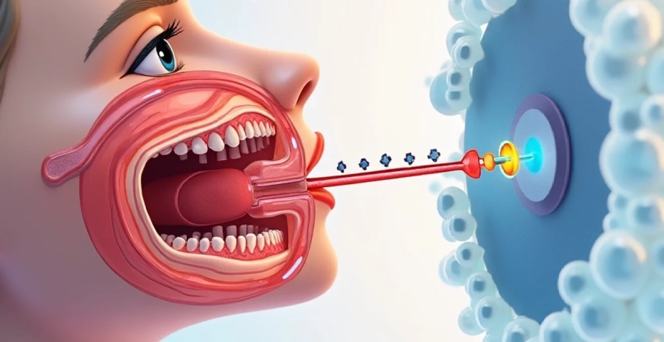
Patulous Eustachian tube (PET) represents one of the most challenging conditions in otolaryngology, affecting thousands of patients worldwide with symptoms that can significantly impact quality of life. Unlike the more common obstructive Eustachian tube dysfunction, PET involves an abnormally patent or open tube that fails to close properly, leading to distressing symptoms such as autophony, aural fullness, and respiratory-synchronous tympanic membrane movement. The complexity of this condition stems from its varied aetiology, ranging from rapid weight loss and hormonal changes to neurological disorders and iatrogenic causes following nasal surgery.
The management of patulous Eustachian tube dysfunction requires a comprehensive understanding of both conservative and surgical interventions, as treatment outcomes can vary significantly between patients. Current therapeutic approaches encompass a broad spectrum of options, from simple postural modifications and nasal preparations to complex surgical reconstructions involving tissue grafting and tube occlusion techniques. The challenge for clinicians lies not only in accurate diagnosis but also in selecting the most appropriate treatment strategy based on individual patient factors, symptom severity, and underlying pathophysiology.
Conservative Non-Surgical management protocols for patulous eustachian tube
Conservative management remains the cornerstone of initial treatment for patients with patulous Eustachian tube dysfunction, particularly those with mild to moderate symptoms or pregnancy-related PET. Non-surgical approaches offer several advantages, including minimal risk of complications, cost-effectiveness, and the potential for symptom resolution without invasive procedures. These conservative strategies focus on addressing the underlying pathophysiology by promoting mucosal swelling, improving tissue hydration, and modifying environmental factors that contribute to tube patency.
The success of conservative management often depends on patient compliance and the systematic application of multiple therapeutic modalities simultaneously. Studies have shown that approximately 40-60% of patients with mild patulous Eustachian tube symptoms can achieve satisfactory improvement through conservative measures alone, making these approaches an essential first-line treatment option.
Nasal decongestant therapy with oxymetazoline and phenylephrine
Topical nasal decongestants represent a paradoxical yet effective treatment approach for patulous Eustachian tube dysfunction. While these medications typically reduce mucosal swelling, their strategic use in PET management involves promoting localised oedema around the Eustachian tube orifice. Oxymetazoline-based preparations applied directly to the nasopharyngeal opening can induce sufficient mucosal swelling to facilitate tube closure, particularly when combined with head-down positioning during application.
Clinical protocols typically involve the application of oxymetazoline 0.05% or phenylephrine 0.25% drops, with patients instructed to lie supine with the head tilted backwards for 10-15 minutes following instillation. The mechanism involves α-adrenergic stimulation leading to vasoconstriction followed by reactive hyperaemia, ultimately resulting in mucosal swelling that can temporarily close the patulous tube.
Postural modification techniques and Head-Down positioning
Postural therapy represents one of the most immediately effective conservative interventions for patulous Eustachian tube symptoms. The physiological basis for this approach lies in the gravitational redistribution of blood flow and the temporary engorgement of mucosal tissues when the head is positioned below heart level. Head-down positioning techniques can provide rapid, albeit temporary, symptom relief by mechanically closing the abnormally patent tube through tissue swelling.
Patients are instructed to assume various positions including lying flat with the head turned to the affected side, placing the head between the knees whilst seated, or performing the Trendelenburg position for 5-10 minutes several times daily. These manoeuvres can be particularly effective during acute symptom episodes, providing immediate relief from autophony and aural fullness.
Weight gain strategies for underweight PET patients
Weight restoration plays a crucial role in managing patulous Eustachian tube dysfunction, particularly in patients who have experienced rapid or significant weight loss. The relationship between body weight and PET symptoms is attributed to the loss of peritonsillar fat pads, including Ostmann’s fat pad, which normally provides structural support for Eustachian tube closure. Structured weight gain programmes can lead to substantial improvement in symptoms, with many patients experiencing complete resolution once optimal weight is restored.
Clinical evidence suggests that patients maintaining a body mass index above certain thresholds experience significantly fewer PET symptoms. Weight gain strategies should involve nutritional counselling, gradual caloric increase, and regular monitoring to ensure healthy weight restoration whilst addressing any underlying metabolic or psychological factors contributing to weight loss.
Saline irrigation and nasal moisturisation protocols
Maintaining optimal mucosal hydration represents a fundamental aspect of conservative PET management. Isotonic saline irrigation helps preserve mucosal integrity whilst promoting the natural healing processes that may contribute to improved Eustachian tube function. Regular nasal irrigation can also help remove inflammatory mediators and allergens that might contribute to mucosal atrophy or dysfunction.
Patients are typically advised to perform saline rinses 2-3 times daily using buffered isotonic solutions, with particular attention to technique and consistency. The addition of humidification therapy, especially during sleep, can further support mucosal health and potentially improve tube closure mechanisms through enhanced tissue hydration.
Minimally invasive surgical interventions for eustachian tube dysfunction
When conservative measures fail to provide adequate symptom relief, minimally invasive surgical interventions offer the next tier of treatment options for patulous Eustachian tube dysfunction. These procedures aim to restore normal tube function whilst minimising surgical morbidity and preserving the potential for future interventions if necessary. Minimally invasive techniques have gained popularity due to their relatively low complication rates, shorter recovery times, and the ability to perform many procedures under local anaesthesia in outpatient settings.
The selection of appropriate minimally invasive procedures depends on several factors, including the severity of symptoms, anatomical considerations, patient age and health status, and previous treatment failures. Success rates for these interventions vary considerably, ranging from 40% to 80% depending on the specific technique employed and patient selection criteria. The reversible nature of many minimally invasive procedures makes them attractive options for patients who may require multiple interventions over time.
Tympanostomy tube insertion and ventilation management
Myringotomy with tympanostomy tube insertion represents one of the most commonly employed surgical interventions for patulous Eustachian tube dysfunction. This procedure addresses the pressure-related symptoms of PET by providing an alternative pathway for middle ear ventilation, effectively bypassing the dysfunctional Eustachian tube. Ventilation tube placement can provide significant relief from autophony and aural fullness, though its effectiveness in eliminating respiratory-synchronous symptoms may be limited.
The procedure involves creating a small incision in the tympanic membrane and inserting a small plastic or metal tube to maintain ventilation. Success rates for symptom improvement range from 50-70%, with many patients experiencing substantial quality of life improvements. However, tubes may require periodic replacement, and some patients may experience increased discomfort or infection rates compared to normal populations.
Cartilage grafting techniques using tragal and conchal cartilage
Autologous cartilage grafting has emerged as a highly effective treatment modality for patulous Eustachian tube dysfunction, offering excellent biocompatibility and long-term stability. Tragal cartilage harvesting provides an ideal donor site due to its proximity to the surgical field and similar tissue characteristics to the structures surrounding the Eustachian tube. The technique involves harvesting small cartilage chips and inserting them submucosally around the tube orifice to provide structural support for closure.
Recent studies have demonstrated success rates of up to 80% for cartilage grafting procedures, with particularly impressive results in patients with well-defined anatomical deficits. The procedure can be performed endoscopically under local anaesthesia, making it suitable for outpatient treatment. Long-term follow-up data suggests that cartilage grafts maintain their structural integrity better than other graft materials, potentially providing more durable results.
Fat plug insertion via transcanal approach
Autologous fat grafting represents another valuable option in the surgical management of patulous Eustachian tube dysfunction. The technique involves harvesting fat tissue, typically from the thigh or abdomen, and injecting it strategically around the Eustachian tube opening to promote closure. Fat plug insertion procedures can be enhanced through the addition of platelet-rich plasma to improve graft survival and integration.
The transcanal approach allows for precise placement of fat grafts whilst minimising surgical trauma and recovery time. Success rates vary from 60-75%, with some patients requiring repeat procedures due to fat resorption over time. The combination of fat grafting with other techniques, such as balloon dilatation, may improve outcomes by optimising the local tissue environment for graft integration.
Eustachian tube balloon dilatation (ETBD) procedures
Balloon dilatation of the Eustachian tube, whilst primarily designed for obstructive dysfunction, has shown promise in selected cases of patulous tube dysfunction when used in combination with other procedures. ETBD techniques can help restore normal tube compliance and may facilitate the success of subsequent grafting procedures by improving local tissue characteristics and vascularity.
The procedure involves inserting a small balloon catheter into the Eustachian tube and inflating it to predetermined pressures for specific durations. When used as part of a combined surgical approach, balloon dilatation may help optimise the tissue environment for subsequent interventions, though its role as a standalone treatment for PET remains controversial among specialists.
Advanced surgical reconstruction methods
For patients with severe, refractory patulous Eustachian tube dysfunction who have failed conservative and minimally invasive treatments, advanced surgical reconstruction methods offer the potential for definitive symptom resolution. These complex procedures require significant surgical expertise and carry higher risks, but they also provide the highest success rates for carefully selected patients. Advanced reconstructive techniques focus on permanently altering the anatomy and physiology of the Eustachian tube to restore normal function whilst maintaining the tube’s essential physiological roles.
The decision to proceed with advanced surgical reconstruction requires careful consideration of multiple factors, including patient age, symptom severity, impact on quality of life, and the presence of comorbid conditions. Success rates for these procedures can exceed 80-90% in experienced hands, but patient selection remains critical for optimal outcomes. The irreversible nature of many advanced techniques necessitates thorough preoperative counselling and realistic expectation setting.
Advanced surgical reconstruction represents the final therapeutic option for patients with debilitating patulous Eustachian tube symptoms, offering the potential for definitive cure but requiring careful patient selection and expert surgical technique.
Surgical techniques in this category include complete Eustachian tube occlusion with simultaneous tympanostomy tube placement, tensor veli palatini muscle transposition, and complex tissue reconstruction using multiple graft materials. The Bluestone technique of tube occlusion using methyl methacrylate-filled catheters has shown excellent long-term results, with success rates approaching 85-90% in suitable candidates. This approach involves accessing the bony portion of the Eustachian tube through an anterior tympanotomy and inserting a catheter filled with biocompatible occlusive material.
Multilayer endoscopic ligation techniques represent another advanced option, combining fat plugging, endoluminal cauterisation, and suture ligation under direct visualisation. These procedures require specialised endoscopic equipment and significant surgical experience but offer the advantage of precise anatomical reconstruction tailored to individual patient anatomy. Success rates for combined multilayer techniques have been reported at 65-85%, with excellent long-term durability in most patients.
Pharmacological treatment approaches and medical management
Pharmaceutical interventions play a supporting role in the comprehensive management of patulous Eustachian tube dysfunction, often serving as adjuvant therapy to other treatment modalities. Medical management strategies focus on addressing underlying inflammatory processes, modifying mucosal characteristics, and managing associated symptoms that contribute to patient discomfort. While pharmacological treatments rarely provide complete symptom resolution as monotherapy, they can significantly enhance the effectiveness of other interventions and improve patient comfort during treatment.
The pharmacological approach to PET management requires an understanding of the complex pathophysiology underlying tube dysfunction and the various factors that may contribute to symptom persistence. Medications must be selected based on individual patient characteristics, comorbid conditions, and potential interactions with other treatments. Long-term medication use requires careful monitoring for side effects and efficacy assessment through regular follow-up evaluations.
Hormonal preparations, particularly oestrogen-containing nasal drops, have shown efficacy in promoting mucosal oedema and facilitating tube closure. The typical preparation involves 25mg of oestrogen (Premarin) in 30ml of normal saline, administered as three drops three times daily to the affected nostril. This treatment can be particularly effective in postmenopausal women or patients with hormonal imbalances contributing to mucosal atrophy.
Saturated potassium iodide solution represents another traditional pharmacological approach, administered as 10 drops in fruit juice three times daily. The mechanism of action involves inducing mucosal swelling through iodide accumulation in secretory glands, though patient tolerance and compliance can be challenging due to the unpalatable taste and potential thyroid effects. Anticholinergic nasal preparations may also provide benefit through their effects on glandular secretions and mucosal characteristics.
Emerging technologies and novel therapeutic interventions
The field of patulous Eustachian tube treatment continues to evolve with the introduction of innovative technologies and novel therapeutic approaches that promise improved outcomes for patients with refractory symptoms. Emerging treatment modalities leverage advances in materials science, bioengineering, and minimally invasive surgical techniques to address the limitations of current therapeutic options. These developments offer hope for patients who have exhausted traditional treatment avenues and may provide more durable solutions with reduced morbidity.
Recent advances in biocompatible materials have led to the development of new injectable substances for Eustachian tube augmentation. Calcium hydroxyapatite injections have shown promise as a semi-permanent treatment option, providing structural support for tube closure whilst maintaining biocompatibility and minimal inflammatory response. The material’s longevity exceeds that of traditional fat grafts, potentially reducing the need for repeat procedures.
Innovation in patulous Eustachian tube treatment continues to drive the development of more effective, less invasive therapeutic options that may revolutionise patient care in the coming years.
Endoscopic surgical techniques continue to advance, with high-definition imaging systems and specialised instrumentation enabling more precise interventions with reduced tissue trauma. Three-dimensional endoscopic systems provide enhanced depth perception during delicate grafting procedures, whilst intraoperative navigation systems help surgeons achieve optimal graft placement and avoid critical anatomical structures.
Tissue engineering approaches represent perhaps the most exciting frontier in PET treatment, with researchers exploring the use of scaffold materials and growth factors to promote natural tissue regeneration around the Eustachian tube. These biological approaches aim to restore normal anatomy and physiology rather than simply providing mechanical occlusion, potentially offering more physiological and durable solutions to tube dysfunction.
Post-treatment monitoring and Long-Term management strategies
Successful management of patulous Eustachian tube dysfunction extends far beyond the initial treatment intervention, requiring comprehensive long-term monitoring and adaptive management strategies to maintain optimal outcomes. Post-treatment surveillance protocols must account for the dynamic nature of PET symptoms, the potential for treatment failure or symptom recurrence, and the need for ongoing patient support and education. Effective long-term management involves regular clinical assessments, objective outcome measurements, and proactive intervention when early signs of treatment failure emerge.
The complexity of post-treatment monitoring stems from the subjective nature of many PET symptoms and the variability in treatment response between individual patients. Standardised outcome measures, including validated symptom questionnaires and objective diagnostic tests, provide essential tools for tracking treatment efficacy and guiding management decisions. Patient education regarding symptom recognition, when to seek medical attention, and realistic expectations for treatment outcomes plays a crucial role in successful long-term management.
Follow-up protocols typically involve initial assessments at 1-2 weeks post-treatment, followed by evaluations at 1, 3, 6, and 12
months post-treatment to monitor for symptom recurrence and treatment efficacy. Long-term surveillance may extend to annual evaluations for patients who achieve stable symptom control, though more frequent monitoring may be necessary for those with complex presentations or multiple comorbidities.
Objective assessment tools play a crucial role in post-treatment monitoring, with tympanometry and nasopharyngeal endoscopy providing valuable insights into treatment success and anatomical changes. Standardised symptom questionnaires such as the Eustachian Tube Dysfunction Questionnaire (ETDQ-7) enable consistent tracking of patient-reported outcomes and facilitate comparison between different treatment modalities. Regular audiometric assessments help monitor for any hearing-related complications or improvements following intervention.
Patient education forms a cornerstone of effective long-term management, empowering individuals to recognise early signs of symptom recurrence and implement appropriate self-management strategies. Patients should be counselled regarding realistic expectations for treatment durability, particularly following procedures with known limitations such as fat grafting or temporary interventions. The importance of maintaining optimal weight, avoiding known precipitating factors, and adhering to prescribed medications must be emphasised during follow-up consultations.
Adaptive management strategies become essential when initial treatments fail to provide sustained relief or when symptoms gradually return over time. Salvage procedures may involve repeat interventions using the same technique, alternative surgical approaches, or combination therapies tailored to individual patient needs. The decision-making process for revision surgery requires careful consideration of previous treatment responses, anatomical changes from prior interventions, and patient preferences regarding additional procedures.
Quality of life assessment represents a critical component of long-term management, as the impact of patulous Eustachian tube dysfunction extends beyond purely medical considerations to affect social interactions, professional activities, and psychological well-being. Regular screening for depression and anxiety, which commonly accompany chronic ear disorders, ensures comprehensive patient care and may identify candidates for additional psychological support or pharmacological intervention.
The establishment of clear communication channels between patients and healthcare providers facilitates prompt intervention when problems arise and helps maintain treatment adherence over time. Emergency contact protocols should be established for patients experiencing severe symptom exacerbations, whilst routine follow-up scheduling ensures continuity of care and early detection of treatment complications.
Successful long-term management of patulous Eustachian tube dysfunction requires a partnership between patient and provider, emphasising education, realistic expectations, and proactive monitoring to achieve optimal outcomes.
Documentation of treatment outcomes and patient responses contributes to the growing body of evidence regarding optimal management strategies for this challenging condition. Systematic data collection enables refinement of treatment protocols and helps identify predictive factors for treatment success, ultimately improving care for future patients with similar presentations.
Future developments in post-treatment monitoring may include telemedicine platforms for remote symptom tracking, smartphone-based applications for patient-reported outcome measures, and advanced imaging techniques for non-invasive assessment of treatment durability. These technological advances promise to enhance the precision and convenience of long-term monitoring whilst reducing the burden on both patients and healthcare systems.