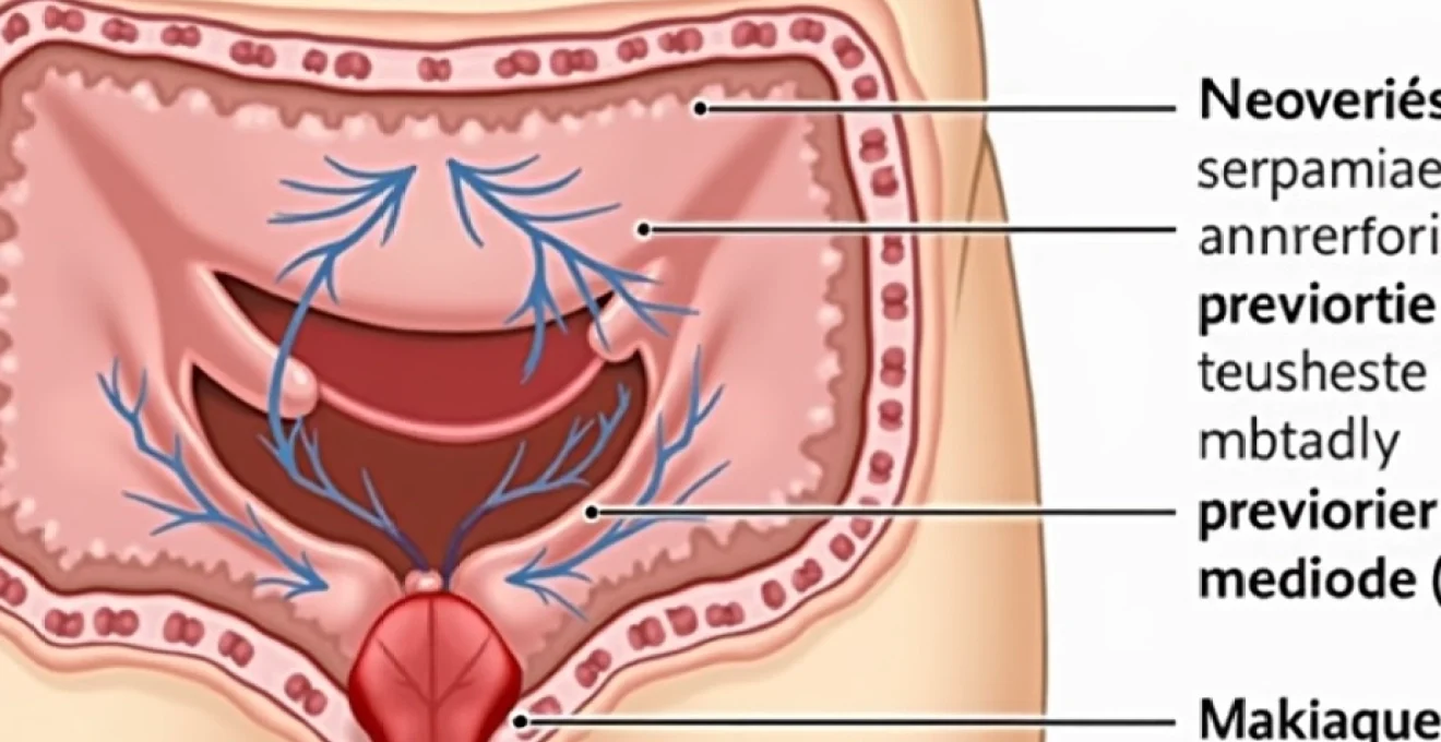
Lateral internal sphincterotomy represents the gold standard surgical intervention for chronic anal fissures that have failed to respond to conservative treatment measures. This carefully executed procedure involves the controlled division of internal anal sphincter fibres to relieve excessive muscle tension that prevents wound healing. With success rates exceeding 95% and relatively low complication rates, sphincterotomy offers definitive relief for patients who have endured weeks or months of debilitating anal pain. The recovery process, whilst generally straightforward, requires careful attention to wound care, bowel management, and progressive functional restoration to achieve optimal outcomes.
Understanding the recovery timeline helps patients prepare for the healing journey ahead and sets realistic expectations for their return to normal activities. The recovery process encompasses immediate post-operative care, wound healing progression, and long-term functional adaptation of the anal sphincter complex.
Understanding lateral internal sphincterotomy surgical technique and indications
The lateral internal sphincterotomy procedure addresses the fundamental pathophysiology underlying chronic anal fissures. When conservative treatments including topical nitrates, calcium channel blockers, and botulinum toxin injections fail to provide lasting relief, surgical intervention becomes necessary. The procedure specifically targets the hypertonic internal anal sphincter, which maintains excessive resting pressure and creates a cycle of ischaemia and delayed healing.
During the operation, surgeons create a precise incision through the internal sphincter fibres at the lateral position, typically at the 3 o’clock or 9 o’clock position when viewed in the lithotomy position. This strategic placement minimises the risk of keyhole deformity whilst achieving adequate pressure reduction. The goal involves decreasing resting anal pressure by approximately 20-30%, sufficient to restore normal blood flow and enable fissure healing.
Division of internal anal sphincter fibres using Lord-Eisenhammer method
The Lord-Eisenhammer technique remains the most widely adopted approach for lateral internal sphincterotomy. This method involves inserting a narrow-bladed scalpel or scissors through a small lateral incision, advancing the instrument to the dentate line, and performing a controlled division of the internal sphincter fibres. The surgeon carefully palpates the groove between the internal and external sphincters to ensure accurate placement and prevent inadvertent damage to the external sphincter complex.
Precise anatomical knowledge proves essential during this procedure, as the internal sphincter extends from the anorectal ring to just below the dentate line. The division typically encompasses the distal two-thirds of the muscle, preserving the proximal fibres that contribute to continence maintenance. Electrocautery may be employed to achieve haemostasis and reduce post-operative bleeding risk.
Chronic anal fissure management through controlled sphincter muscle release
Chronic anal fissures develop when acute tears fail to heal within 6-8 weeks, often progressing to expose the underlying internal sphincter muscle. The exposed muscle triggers protective spasm reflexes that further compromise blood supply and perpetuate the non-healing cycle. These fissures frequently develop characteristic features including sentinel skin tags , hypertrophied anal papillae, and indurated margins with visible muscle fibres at the fissure base.
The controlled release achieved through sphincterotomy interrupts this pathological cycle by reducing sphincter tone to physiological levels. This intervention restores adequate perfusion to the anoderm, enabling the body’s natural healing mechanisms to repair the chronic wound. Studies demonstrate that properly performed sphincterotomy achieves fissure healing in over 95% of patients within 6-8 weeks post-operatively.
Open versus closed sphincterotomy approaches in colorectal surgery
Surgeons may employ either open or closed techniques for lateral internal sphincterotomy, each offering distinct advantages depending on patient factors and surgeon preference. The open technique involves direct visualisation of the sphincter fibres through a small radial incision, allowing precise identification and division of the muscle under direct vision. This approach facilitates accurate depth control and enables concurrent treatment of associated pathology such as sentinel tags or hypertrophied papillae.
The closed technique utilises palpation to identify the intersphincteric groove and performs blind division of the internal sphincter through a small stab incision. Whilst this method offers cosmetic advantages and potentially faster healing, it requires greater technical expertise to ensure adequate sphincter division whilst avoiding external sphincter injury. Both techniques demonstrate comparable efficacy rates when performed by experienced colorectal surgeons.
Patient selection criteria for lateral internal sphincterotomy procedures
Appropriate patient selection remains crucial for achieving optimal outcomes whilst minimising complication risks. Ideal candidates include individuals with chronic posterior or anterior anal fissures that have failed 8-12 weeks of maximal medical therapy. Pre-operative assessment should evaluate sphincter function through digital examination and may include anal manometry in selected cases to assess baseline pressures.
Certain patient populations require careful consideration before proceeding with sphincterotomy. Women with obstetric trauma history, elderly patients with diminished sphincter function, and individuals with inflammatory bowel disease may face increased incontinence risks. In such cases, alternative procedures including advancement flaps or fissurectomy with botulinum toxin injection may prove more appropriate treatment options.
Immediate Post-Operative recovery timeline and expectations
The immediate post-operative period following lateral internal sphincterotomy sets the foundation for successful long-term recovery. Most procedures are performed as day-case surgery under general or regional anaesthesia, allowing patients to return home within 4-6 hours post-operatively. The initial recovery phase focuses on pain management, wound care initiation, and early mobilisation to prevent complications such as urinary retention or deep vein thrombosis.
Patients typically experience immediate relief from the chronic fissure pain that prompted surgical intervention, though some degree of post-operative discomfort should be anticipated. The surgical site may be tender for several days, and the first bowel movement often generates anxiety despite being generally less painful than pre-operative defaecation. Establishing realistic expectations during this period helps patients navigate the early recovery challenges whilst maintaining confidence in the treatment decision.
First 24-48 hours: acute pain management and wound assessment
Pain management during the initial 24-48 hours post-sphincterotomy typically involves a multimodal approach combining oral analgesics with topical preparations. Paracetamol and non-steroidal anti-inflammatory drugs form the foundation of pain control, whilst short-term opioid use may be necessary for breakthrough pain. Topical anaesthetic preparations containing lidocaine can provide additional relief when applied to the perianal area.
Wound assessment during this period focuses on monitoring for excessive bleeding, signs of haematoma formation, or early infection indicators. Minor bleeding from the surgical site is expected and typically resolves spontaneously within 48 hours. Patients should be advised to observe for concerning symptoms including persistent heavy bleeding, severe pain unresponsive to prescribed medications, or signs of systemic infection such as fever or malaise.
Week one recovery: bowel movement normalisation and dietary modifications
The first week post-sphincterotomy represents a critical period for establishing healthy bowel patterns that support continued healing. Dietary modifications play a central role during this phase, with emphasis placed on maintaining soft, well-formed stools that minimise trauma to the healing surgical site. High-fibre foods including fruits, vegetables, whole grains, and legumes should be gradually introduced alongside adequate fluid intake of 2-3 litres daily.
Stool softeners such as docusate sodium or osmotic laxatives like macrogol may be prescribed to prevent constipation and reduce straining during defaecation. The goal involves achieving effortless bowel movements that do not compromise the healing process or cause recurrent trauma to the anal canal. Patients should be encouraged to respond promptly to defaecatory urges and avoid prolonged straining attempts that could increase anal pressure.
Two-week mark: tissue healing progress and suture dissolution
By the two-week milestone, significant tissue healing should be evident with reduced pain levels and improved comfort during bowel movements. Any absorbable sutures placed during the procedure typically begin dissolving during this period, though complete dissolution may take 3-4 weeks. The surgical site gradually develops mature scar tissue that provides structural integrity whilst maintaining the desired reduction in sphincter tone.
Patients may notice gradual improvement in their ability to discriminate between gas and solid stool, indicating restoration of normal sphincter function and sensation. Some individuals experience temporary alterations in continence during this healing phase, which usually resolve as swelling subsides and tissues adapt to the new anatomy. Regular sitz baths in warm water continue to provide symptomatic relief and promote healing through improved local blood flow.
Month one milestone: sphincter function adaptation and continence evaluation
The four-week mark represents a significant milestone in sphincterotomy recovery, with most patients achieving substantial symptom resolution and functional improvement. By this stage, the original anal fissure should demonstrate complete healing, and pain levels should be minimal or absent. Sphincter function typically stabilises during this period, with patients regaining normal discriminatory ability and continence control.
Continence evaluation at one month post-operatively helps identify any persistent functional deficits that may require additional intervention. The vast majority of patients maintain perfect continence, whilst a small percentage may experience minor gas incontinence or occasional soiling episodes. These symptoms often improve with time and may respond to pelvic floor physiotherapy or dietary modifications to optimise stool consistency.
Managing Post-Sphincterotomy complications during recovery
Whilst lateral internal sphincterotomy demonstrates excellent safety profiles, potential complications may arise during the recovery period requiring prompt recognition and appropriate management. Early identification of complications enables timely intervention and prevents progression to more serious problems that could compromise long-term outcomes. Understanding the spectrum of potential complications helps patients and healthcare providers maintain vigilance during the critical healing period.
The most commonly encountered complications include temporary incontinence, wound infections, haematoma formation, and recurrent fissuring. Each complication presents unique challenges and requires specific management strategies tailored to the individual patient’s circumstances and severity of symptoms. Proactive monitoring and early intervention significantly improve outcomes for patients who develop post-operative complications.
Faecal incontinence risk assessment and kegel exercise protocols
Faecal incontinence represents the most concerning potential complication following sphincterotomy, though significant incontinence occurs in fewer than 5% of patients when performed by experienced surgeons. Minor incontinence to gas or liquid stool may affect 10-15% of patients temporarily, typically resolving within 2-3 months as tissues heal and adapt. Risk factors for incontinence include advanced age, female gender, previous obstetric trauma, and pre-existing sphincter weakness.
Kegel exercises and pelvic floor strengthening protocols prove beneficial for patients experiencing temporary incontinence symptoms. These exercises involve rhythmic contraction and relaxation of the pelvic floor muscles, performed 3-4 times daily in sets of 10-15 repetitions. Biofeedback therapy may enhance exercise effectiveness by providing visual or auditory feedback regarding muscle activation patterns. Most patients notice gradual improvement in continence control within 4-6 weeks of initiating structured exercise programmes.
Wound infection prevention using metronidazole and topical antiseptics
Wound infection prevention relies primarily on meticulous hygiene practices and appropriate antibiotic prophylaxis when indicated. The perianal location of the surgical site predisposes to bacterial contamination from faecal matter, necessitating careful attention to cleansing routines and protective measures. Gentle cleansing with warm water after each bowel movement, followed by thorough drying, forms the cornerstone of infection prevention strategies.
Metronidazole may be prescribed prophylactically for high-risk patients or those developing early signs of infection such as increased pain, purulent discharge, or erythema extending beyond the immediate surgical site. Topical antiseptic preparations containing povidone-iodine or chlorhexidine can provide additional protection when applied to the perianal area after cleansing. Signs of serious infection requiring urgent medical attention include fever, spreading cellulitis, or abscess formation.
Haematoma formation management and sitz bath therapy implementation
Haematoma formation occurs in approximately 2-3% of patients following sphincterotomy, typically presenting as a tender, fluctuant mass adjacent to the surgical site. Small haematomas often resolve spontaneously with conservative management, whilst larger collections may require drainage to prevent complications such as infection or delayed healing. Early recognition enables appropriate triage between conservative and surgical management approaches.
Sitz bath therapy provides effective management for small haematomas whilst promoting overall healing and comfort. Patients should immerse the perineal area in warm water (approximately 37-40°C) for 15-20 minutes, 2-3 times daily, particularly after bowel movements. The warm water promotes vasodilation, enhances blood flow, and facilitates haematoma resolution through improved lymphatic drainage. Adding Epsom salts to the bath water may provide additional anti-inflammatory benefits.
Recurrent anal fissure prevention through stool softener regimens
Preventing recurrent anal fissure formation requires ongoing attention to bowel habits and stool consistency maintenance even after successful sphincterotomy. Whilst recurrence rates following properly performed sphincterotomy remain low (less than 5%), patients must maintain vigilance regarding factors that contributed to their original fissure development. Chronic constipation represents the most significant risk factor for recurrence and requires long-term management strategies.
Prophylactic stool softener regimens may be recommended for patients with recurrent constipation history or those at high risk for developing hard stools. Osmotic laxatives such as polyethylene glycol provide effective long-term management with minimal side effects when used appropriately. Dietary counselling emphasising adequate fibre intake (25-35 grams daily) and proper hydration remains fundamental to preventing recurrent fissuring and maintaining optimal bowel function.
Long-term recovery outcomes and functional assessment
Long-term outcomes following lateral internal sphincterotomy demonstrate excellent success rates with sustained symptom relief and high patient satisfaction scores. Studies with follow-up periods extending beyond five years consistently report healing rates exceeding 95% with minimal recurrence and preserved quality of life. The procedure’s durability stems from its ability to address the underlying pathophysiology of chronic anal fissures whilst maintaining functional sphincter integrity in the vast majority of patients.
Functional assessment at six months and one year post-operatively provides valuable insights into the procedure’s long-term efficacy and identifies any delayed complications requiring intervention. Most patients report complete resolution of pain symptoms and return to normal activities without dietary restrictions or ongoing medication requirements. The minority of patients experiencing persistent symptoms may benefit from additional interventions or specialist evaluation to identify contributing factors.
Quality of life improvements following successful sphincterotomy extend beyond simple pain relief to encompass psychological and social benefits. Many patients describe renewed confidence in their ability to engage in work, travel, and recreational activities without fear of debilitating pain episodes. The restoration of normal bowel function eliminates the anxiety and avoidance behaviours that often develop in response to chronic anal fissure symptoms, enabling patients to resume their preferred lifestyle patterns.
Long-term continence outcomes remain favourable, with serious incontinence complications affecting fewer than 2% of patients when surgery is performed by experienced colorectal surgeons. Minor gas incontinence or occasional seepage may persist in 3-5% of patients but rarely interferes significantly with daily activities or quality of life. These symptoms often respond well to conservative management including dietary modifications, pelvic floor exercises, and topical barrier preparations.
Comparative recovery analysis: sphincterotomy versus alternative treatments
When comparing recovery trajectories between lateral internal sphincterotomy and alternative treatment modalities, several key differences emerge that influence treatment selection and patient counselling. Sphincterotomy typically offers the most rapid and complete symptom resolution, with many patients experiencing immediate relief from chronic pain. Alternative treatments such as topical nitrates, calcium channel blockers, or botulinum toxin injections may require multiple treatment cycles and demonstrate lower success rates for chronic fissures.
Recovery time following sphincterotomy generally proves shorter and more predictable compared to non-surgical alternatives. Whilst medical therapies may require 8-12 weeks of treatment with uncertain outcomes, sphincterotomy patients typically achieve complete healing within 4-6 weeks post-operatively. The definitive nature of surgical treatment eliminates the need for ongoing medication compliance and reduces the likelihood of treatment failure or symptom recurrence.
The economic considerations of different treatment approaches also favour sphincterotomy for appropriate candidates. Whilst the initial cost may exceed that of medical therapies, the reduced need for multiple consultations, repeated prescriptions, and treatment failures often results in lower overall healthcare expenditure. Patients benefit from shorter time away from work and reduced impact on productivity compared to prolonged medical management cycles.
However, certain patient populations may experience better recovery outcomes with alternative treatments. Young women planning pregnancy, patients with pre-existing continence issues, or those with inflammatory bowel disease may benefit from the reversible nature of medical therapies or less invasive procedures such as advancement flaps. The recovery from these alternatives may involve longer healing periods but offers different risk-benefit profiles that suit specific clinical scenarios.
Optimising recovery through evidence-based rehabilitation protocols
Evidence-based rehabilitation protocols following lateral internal sphincterotomy focus on supporting natural healing processes whilst minimising complications and optimising functional outcomes. These protocols integrate nutritional guidance, physical therapy principles, and behavioural modifications to create comprehensive recovery frameworks. Research demonstrates that structured rehabilitation approaches can reduce recovery times by 20-30% compared to standard care protocols.
The foundation of effective rehabilitation rests on understanding the physiological changes occurring during sphincter healing and adaptation. As the divided internal sphincter fibres heal, scar tissue formation must balance structural integrity with maintained pressure reduction. Excessive scar formation can negate the benefits of sphincterotomy, whilst inadequate healing may predispose to recurrent symptoms or complications.
Progressive mobilisation protocols encourage early return to normal activities whilst avoiding excessive strain on the healing tissues. Patients typically begin with gentle walking within 24 hours post-operatively, progressing to normal daily activities within one week. Heavy lifting restrictions (greater than 10 kg) remain in place for 2-3 weeks to prevent increased intra-abdominal pressure that could compromise healing or cause bleeding complications.
Nutritional rehabilitation emphasises maintaining optimal bowel function through strategic dietary modifications that support both healing and long-term fissure prevention. The protocol incorporates graduated fibre introduction, beginning with soluble fibres from sources such as oats, beans, and fruits, followed by insoluble fibres from vegetables and whole grains. This approach minimises the risk of excessive gas production or loose stools that could compromise early healing.
Hydration strategies form an integral component of rehabilitation protocols, with recommended fluid intake of 35-40 ml per kilogram of body weight daily. Adequate hydration supports stool softening, promotes tissue healing through enhanced perfusion, and facilitates the elimination of metabolic waste products. Patients learn to monitor urine colour as a practical indicator of hydration status, aiming for pale yellow throughout the recovery period.
Psychological support elements address the anxiety and apprehension many patients experience regarding bowel movements following anal surgery. Cognitive behavioural techniques help patients overcome avoidance behaviours and develop confidence in their recovering function. Education about normal healing progression and expected sensations reduces anxiety and promotes adherence to rehabilitation protocols.
Regular follow-up assessments enable healthcare providers to monitor recovery progress and adjust rehabilitation protocols based on individual response patterns. These assessments typically occur at 2 weeks, 6 weeks, and 3 months post-operatively, focusing on pain levels, functional capacity, and quality of life measures. Early identification of suboptimal healing allows for prompt intervention before complications develop into more serious problems.
Long-term maintenance strategies extend beyond the acute recovery period to prevent recurrent fissure development and maintain optimal anal health. These strategies emphasise sustainable lifestyle modifications rather than short-term interventions, recognising that chronic anal fissures often reflect underlying predisposing factors requiring ongoing management. Patient education programs demonstrate superior outcomes when they address both immediate recovery needs and long-term prevention strategies within a unified framework.