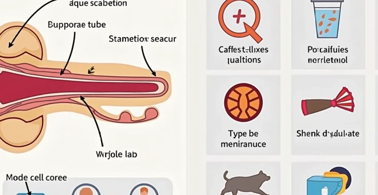
The distinctive popping sensation you experience when swallowing is one of the most common yet fascinating physiological phenomena involving your auditory system. This occurrence stems from the intricate relationship between your middle ear, the Eustachian tube, and the complex pressure regulation mechanisms that maintain optimal hearing function. Understanding why your ears pop during deglutition requires exploring the sophisticated anatomical structures and neurological processes that work seamlessly to protect your hearing whilst adapting to environmental pressure changes.
The popping sound represents your body’s automatic response to pressure differentials, demonstrating the remarkable engineering of the human ear. This natural mechanism ensures that the delicate structures within your middle ear remain functional and protected from potential damage caused by pressure imbalances. Medical professionals recognise this as a crucial aspect of auditory health maintenance.
Eustachian tube anatomy and physiological function
The Eustachian tube serves as the primary anatomical structure responsible for the characteristic ear popping sensation during swallowing. This narrow passageway, measuring approximately 31-38 millimetres in adults, connects your middle ear cavity to the nasopharynx, creating a vital communication channel between these two distinct anatomical regions. The tube’s strategic positioning allows for efficient pressure equalisation whilst maintaining the sterile environment of the middle ear space.
Cartilaginous and osseous portions of the eustachian tube
The Eustachian tube comprises two distinct anatomical segments that work in harmony to facilitate pressure regulation. The osseous portion, constituting approximately one-third of the tube’s total length, extends from the middle ear through the temporal bone. This bony segment remains relatively rigid and serves as the stable foundation for the tube’s mechanical function. The cartilaginous portion, representing the remaining two-thirds, extends towards the nasopharynx and demonstrates remarkable flexibility during the opening and closing mechanisms.
The junction between these two portions creates a natural narrowing known as the isthmus, which acts as a functional valve controlling airflow between the middle ear and nasopharynx. This anatomical design ensures that the middle ear remains sealed under normal circumstances whilst allowing for rapid pressure equalisation when necessary. The cartilaginous framework consists of elastic cartilage that can deform and return to its original shape, enabling the repetitive opening and closing actions required for proper function.
Tensor veli palatini and levator veli palatini muscle actions
Two primary muscles orchestrate the opening mechanism of the Eustachian tube during swallowing activities. The tensor veli palatini muscle, innervated by the mandibular division of the trigeminal nerve, provides the primary force for tube opening. When this muscle contracts during swallowing, it exerts tension on the cartilaginous portion of the tube, creating the mechanical opening that allows pressure equalisation to occur.
The levator veli palatini muscle, controlled by the vagus nerve, provides additional support for tube function through its influence on the soft palate elevation. This coordinated muscular activity ensures that the tube opening occurs at precisely the right moment during the swallowing sequence. The synchronisation between these muscles represents a sophisticated neurological control mechanism that has evolved to protect your hearing whilst maintaining respiratory and digestive functions.
Mucociliary clearance mechanisms in tube ventilation
The internal surface of the Eustachian tube features specialised ciliated epithelium that facilitates continuous clearance of secretions and debris. These microscopic hair-like structures beat in coordinated waves, creating a conveyor belt effect that transports mucus and potential pathogens away from the middle ear towards the nasopharynx. This mucociliary escalator operates continuously, even when the tube remains closed, ensuring optimal hygiene within the middle ear space.
The effectiveness of this clearance mechanism directly influences your susceptibility to ear infections and tube dysfunction. When the mucociliary system becomes compromised due to inflammation, infection, or environmental factors, the resulting accumulation of secretions can interfere with normal tube function. This interference may manifest as persistent ear popping, fullness sensations, or reduced hearing acuity, highlighting the importance of maintaining healthy mucociliary function.
Epithelial lining and goblet cell secretions
The epithelial lining of the Eustachian tube contains numerous goblet cells that produce protective mucus secretions. These specialised cells respond to various stimuli, including mechanical irritation, chemical exposure, and inflammatory mediators, by increasing or decreasing mucus production accordingly. The composition of these secretions includes antimicrobial peptides, immunoglobulins, and other protective substances that help maintain the sterile environment of the middle ear.
The balance between mucus production and clearance determines the overall health of the Eustachian tube system. Excessive secretion production can lead to tube blockage and dysfunction, whilst insufficient production may compromise the protective barrier function. Understanding this delicate balance helps explain why certain conditions, such as allergies or upper respiratory infections, can significantly impact ear popping sensations and overall auditory function.
Barometric pressure equalisation during deglutition
The fundamental reason behind ear popping during swallowing lies in the sophisticated pressure equalisation system that maintains optimal middle ear function. Your middle ear cavity normally contains air at atmospheric pressure, but various factors can create pressure differentials that require active correction. When you swallow, the coordinated muscle contractions temporarily open the Eustachian tube, allowing rapid pressure equalisation between the middle ear and the external environment.
This pressure equalisation process occurs within milliseconds of tube opening, demonstrating the efficiency of your auditory system’s protective mechanisms. The popping sound you hear represents the sudden movement of the tympanic membrane as pressures equalise across its surface. This membrane movement creates acoustic vibrations that your inner ear interprets as the characteristic popping sensation, confirming that the equalisation process has occurred successfully.
Valsalva manoeuvre effects on middle ear pressure
The Valsalva manoeuvre represents a deliberate technique for forcing Eustachian tube opening when natural mechanisms prove insufficient. By closing your mouth, pinching your nostrils, and gently exhaling, you create positive pressure within your respiratory system that can overcome minor tube blockages. This increased pressure forces air through the Eustachian tube into the middle ear, often accompanied by the familiar popping sensation.
However, excessive force during the Valsalva manoeuvre can potentially damage delicate middle ear structures, including the tympanic membrane and ossicular chain. Medical professionals recommend using gentle pressure and discontinuing the technique if discomfort occurs. The effectiveness of this manoeuvre varies among individuals, depending on factors such as tube anatomy, inflammation levels, and underlying medical conditions that may affect tube function.
Toynbee phenomenon and passive tube opening
The Toynbee phenomenon describes the passive opening of the Eustachian tube that occurs during swallowing with a closed nose and mouth. This technique combines the natural swallowing mechanism with modified pressure dynamics to encourage tube opening when conventional swallowing proves inadequate. The phenomenon demonstrates the adaptability of your auditory system’s pressure regulation mechanisms under various conditions.
This passive opening technique proves particularly useful during air travel or diving activities when external pressure changes challenge your natural equalisation abilities. The combination of muscular contraction from swallowing and the pressure modifications created by nasal occlusion often succeeds in opening stubborn tubes that resist conventional equalisation attempts. Understanding this phenomenon provides valuable insight into managing ear discomfort during pressure-challenging activities.
Atmospheric pressure gradients and tympanic membrane response
The tympanic membrane serves as a sensitive barometer, responding immediately to pressure differences between the middle ear and external environment. When atmospheric pressure changes occur faster than your Eustachian tube can equalise, the membrane deflects inward or outward, creating tension that you perceive as ear fullness or discomfort. This deflection can affect hearing acuity and comfort until equalisation occurs through swallowing or other tube-opening mechanisms.
The membrane’s response to pressure gradients follows predictable patterns based on the magnitude and rate of pressure change. Rapid pressure changes, such as those experienced during aircraft descent, create more pronounced membrane deflection and associated discomfort. Understanding these response patterns helps explain why certain activities trigger more intense ear popping sensations and why some individuals experience greater sensitivity to pressure changes than others.
Politzer method for artificial tube inflation
The Politzer method represents a clinical technique for artificially inflating the Eustachian tube when natural mechanisms fail to provide adequate pressure equalisation. This procedure involves introducing positive pressure into the nasal cavity whilst the patient swallows, forcing air through the tube into the middle ear space. Medical professionals utilise this technique for diagnostic purposes and therapeutic intervention in cases of persistent tube dysfunction.
The success of the Politzer method depends on proper technique execution and the underlying condition affecting tube function. While effective for many patients, this approach requires professional supervision to prevent complications such as excessive pressure application or introduction of infectious material into the middle ear. The method provides valuable information about tube patency and function whilst offering therapeutic benefits for select patients experiencing persistent ear pressure issues.
Neurological control mechanisms of ear popping
The neurological orchestration of ear popping involves sophisticated coordination between multiple cranial nerves and central nervous system pathways. The trigeminal nerve (cranial nerve V) provides motor innervation to the tensor veli palatini muscle, whilst the vagus nerve (cranial nerve X) controls the levator veli palatini muscle. This dual innervation ensures redundancy in the system and allows for fine-tuned control of tube opening mechanics.
The swallowing reflex, mediated by the medulla oblongata, coordinates the precise timing of muscle contractions required for effective Eustachian tube opening. This central control mechanism integrates sensory information from the oral cavity, pharynx, and middle ear to determine the appropriate response to pressure differentials. The sophistication of this neurological control system explains why ear popping occurs so reliably during swallowing activities, even without conscious awareness of the process.
Sensory feedback from baroreceptors within the middle ear provides continuous information about pressure status to the central nervous system. When these receptors detect pressure imbalances, they trigger reflexive responses that promote swallowing or other tube-opening behaviours. This feedback loop ensures that pressure equalisation occurs automatically in response to changing environmental conditions, protecting your hearing whilst maintaining comfort and optimal auditory function.
The neurological precision required for proper Eustachian tube function demonstrates the evolutionary importance of maintaining optimal middle ear pressure for survival and communication abilities.
Pathological conditions affecting eustachian tube dysfunction
Various pathological conditions can disrupt the normal function of the Eustachian tube, leading to altered ear popping sensations or complete absence of pressure equalisation. These conditions range from acute inflammatory processes to chronic structural abnormalities, each presenting unique challenges for maintaining proper middle ear function. Understanding these pathological states provides insight into when ear popping changes might indicate underlying medical issues requiring professional evaluation.
Allergic rhinitis and mucosal oedema impact
Allergic rhinitis significantly impacts Eustachian tube function through the development of mucosal oedema and increased secretion production. When allergens trigger inflammatory responses in the nasal cavity and nasopharynx, the resulting tissue swelling can compress or obstruct the tube opening, preventing normal pressure equalisation. This obstruction often manifests as reduced ear popping effectiveness during swallowing, accompanied by feelings of ear fullness or mild hearing impairment.
The inflammatory cascade associated with allergic reactions also increases goblet cell activity, leading to excessive mucus production within the tube lumen. This increased secretion volume can overwhelm the natural mucociliary clearance mechanisms, resulting in tube blockage and dysfunction. Patients with seasonal allergies often report cyclical patterns of ear symptoms that correlate with allergen exposure levels and environmental factors such as pollen counts.
Chronic allergic inflammation can lead to permanent structural changes within the Eustachian tube, including mucosal thickening and reduced tube compliance. These long-term adaptations may require ongoing medical management to maintain adequate tube function and prevent complications such as chronic otitis media or hearing loss. The relationship between allergic rhinitis and tube dysfunction underscores the importance of comprehensive allergy management in maintaining optimal auditory health.
Adenoid hypertrophy and nasopharyngeal obstruction
Adenoid hypertrophy represents a common cause of Eustachian tube dysfunction, particularly in paediatric populations. The enlarged adenoid tissue can physically obstruct the nasopharyngeal opening of the tube, preventing normal pressure equalisation and creating a predisposition to middle ear infections. Children with significant adenoid enlargement often experience persistent ear popping difficulties, frequent ear infections, and associated hearing impairments that can affect speech and language development.
The degree of obstruction caused by adenoid hypertrophy varies depending on the size and location of the enlarged tissue. Complete obstruction eliminates the ability to equalise pressure through swallowing, whilst partial obstruction may allow limited function under certain conditions. This variability explains why some children experience intermittent ear symptoms that worsen during upper respiratory infections when additional inflammation compounds the existing obstruction.
Patulous eustachian tube syndrome manifestations
Patulous Eustachian tube syndrome represents the opposite extreme of tube dysfunction, characterised by abnormal tube patency rather than obstruction. Patients with this condition experience constant or intermittent tube opening, leading to unusual auditory symptoms including autophony (hearing one’s own voice amplified), respiratory synchronous ear sensations, and altered ear popping patterns. The condition can significantly impact quality of life and may require specialised treatment approaches.
The aetiology of patulous tube syndrome includes factors such as rapid weight loss, pregnancy, certain medications, and anatomical variations that affect tube mechanics. Understanding this condition highlights the importance of maintaining appropriate tube function – neither excessive closure nor inappropriate opening provides optimal middle ear protection and pressure regulation. Diagnosis often requires specialised testing and careful clinical evaluation to differentiate from other forms of tube dysfunction.
Chronic otitis media with effusion complications
Chronic otitis media with effusion develops when persistent Eustachian tube dysfunction allows fluid accumulation within the middle ear space. This fluid accumulation interferes with normal tympanic membrane movement and ossicular chain function, resulting in conductive hearing loss and altered pressure sensation. Patients with this condition may experience reduced effectiveness of ear popping during swallowing due to the damping effect of fluid on membrane movement.
The presence of middle ear effusion creates a complex pathophysiological state where normal pressure equalisation mechanisms become compromised. The fluid acts as a barrier to pressure transmission whilst simultaneously providing a medium for bacterial growth and biofilm formation. This environment perpetuates the cycle of inflammation and dysfunction, often requiring active medical intervention to restore normal tube function and prevent long-term hearing complications.
Clinical assessment and diagnostic procedures
Clinical evaluation of Eustachian tube function requires a comprehensive approach that combines patient history, physical examination, and specialised diagnostic testing. Healthcare professionals assess tube function through various methods, including tympanometry, acoustic reflectometry, and direct visualisation techniques. These assessments provide objective measurements of tube patency, pressure regulation capability, and overall middle ear health status.
Tympanometry represents the gold standard for evaluating middle ear pressure and Eustachian tube function. This test measures tympanic membrane compliance across a range of applied pressures, providing information about tube patency and middle ear status. Type A tympanograms indicate normal function, whilst Type B and C patterns suggest various degrees of dysfunction requiring further evaluation and potential intervention. The test’s non-invasive nature makes it suitable for patients of all ages and clinical conditions.
Advanced diagnostic techniques include tubomanometry, which directly measures pressure changes within the Eustachian tube during swallowing activities. This specialised testing provides detailed information about tube opening dynamics, duration of patency, and pressure equalisation efficiency. Such comprehensive evaluation helps clinicians develop targeted treatment strategies based on the specific nature and severity of tube dysfunction present in individual patients.
Accurate diagnosis of Eustachian tube dysfunction requires careful correlation between subjective symptoms, objective findings, and functional assessment results to ensure appropriate treatment selection.
Preventative measures and therapeutic interventions
Maintaining optimal Eustachian tube function involves a combination of lifestyle modifications, environmental considerations, and targeted therapeutic interventions when necessary. Preventative strategies focus on reducing factors that contribute to tube inflammation or dysfunction, including allergen avoidance, proper nasal hygiene, and appropriate management of upper respiratory conditions. These measures can significantly reduce the frequency and severity of ear pressure problems whilst promoting overall auditory health.
Simple techniques such as proper swallowing mechanics, nasal saline irrigation, and controlled pressure equalisation exercises can help maintain tube patency and function. Regular practice of these techniques proves particularly beneficial for individuals frequently
exposed to altitude changes, such as frequent air travellers or professional divers. Educational approaches help individuals recognise early signs of tube dysfunction and implement appropriate self-care measures before symptoms become problematic.
Therapeutic interventions for Eustachian tube dysfunction range from conservative medical management to surgical procedures, depending on the underlying cause and severity of symptoms. Topical nasal decongestants can provide temporary relief by reducing mucosal swelling and improving tube patency, whilst corticosteroid preparations address inflammatory components of dysfunction. Antihistamines prove beneficial for patients with allergic contributions to their tube problems, offering both symptom relief and prevention of future episodes.
For patients with persistent dysfunction despite medical management, surgical options include myringotomy with tube placement, balloon dilation of the Eustachian tube, and adenoidectomy when indicated. These procedures aim to restore normal pressure regulation and prevent complications such as chronic otitis media or permanent hearing loss. The selection of appropriate therapeutic approaches requires careful consideration of patient-specific factors, including age, underlying medical conditions, and individual response to conservative treatments.
Innovative treatment modalities continue to emerge as our understanding of Eustachian tube physiology advances. Techniques such as controlled pressure therapy and specialised physiotherapy exercises show promise for managing certain types of tube dysfunction. These developments offer hope for patients with complex or refractory conditions who may not respond adequately to conventional treatment approaches, highlighting the ongoing evolution of therapeutic options in this specialised field.
Effective management of Eustachian tube dysfunction requires a personalised approach that considers both the underlying pathophysiology and individual patient factors to achieve optimal outcomes whilst minimising treatment-related complications.