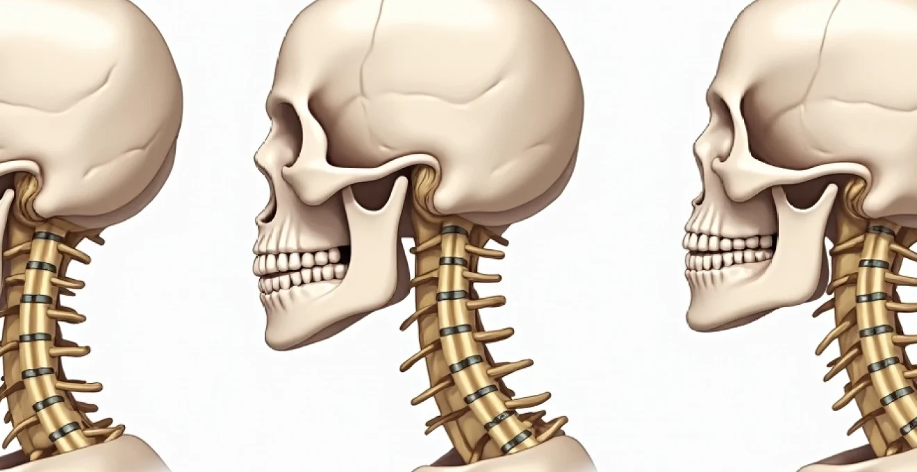
Reverse cervical lordosis represents a significant deviation from the spine’s natural architecture, where the neck’s characteristic inward curve transforms into a problematic outward bend or completely flattens. This condition affects millions of individuals worldwide, often developing silently over years of poor postural habits, degenerative changes, or traumatic injuries. Understanding the intricacies of cervical spine alignment becomes crucial when considering that the neck serves as the vital bridge between the brain and body, supporting approximately 10-12 pounds of head weight while facilitating complex neurological communications.
The transformation from healthy cervical lordosis to its reverse counterpart creates a cascade of biomechanical compensations throughout the entire spinal column. Modern lifestyle factors, including prolonged computer use and smartphone dependency, have dramatically increased the prevalence of cervical alignment disorders. Research indicates that forward head posture, a primary contributor to reverse cervical lordosis, affects up to 66% of the population , making this condition a contemporary epidemic that demands comprehensive understanding and proactive management strategies.
Cervical spine anatomy and normal lordotic curvature mechanics
The cervical spine’s architectural design represents one of nature’s most sophisticated engineering solutions, balancing mobility, stability, and neurological protection within seven vertebral segments. A healthy cervical lordosis measures between 31 and 40 degrees, creating the characteristic C-shaped curve that faces forward toward the body’s centre. This curvature functions like a coiled spring, distributing mechanical forces efficiently whilst maintaining optimal positioning for the head’s centre of mass directly above the shoulders.
C1-C7 vertebral structure and physiological alignment patterns
Each cervical vertebra possesses unique anatomical features that contribute to the spine’s overall lordotic configuration. The atlas (C1) and axis (C2) form a specialised joint complex allowing approximately 50% of total cervical rotation, whilst the remaining five vertebrae (C3-C7) provide the primary lordotic curvature through their wedge-shaped bodies. The vertebral bodies progressively increase in size from C3 to C7 , creating a natural tendency toward lordotic alignment when proper muscle balance exists.
Intervertebral disc height distribution plays a crucial role in maintaining physiological lordosis, with the C5-C6 and C6-C7 levels typically exhibiting the greatest disc thickness. These lower cervical segments bear the highest mechanical loads and demonstrate the most pronounced lordotic angulation. The facet joints orient at approximately 45 degrees to the horizontal plane, facilitating smooth flexion-extension movements whilst providing stability against excessive rotation and lateral bending forces.
Atlas-axis complex role in cervical lordosis maintenance
The atlantooccipital and atlantoaxial joints function as the cervical spine’s primary motion centres, contributing significantly to overall lordotic positioning. The atlas lacks a vertebral body, instead forming a ring-like structure that articulates with the occiput above and axis below. This unique configuration allows the head to achieve fine positional adjustments whilst the axis provides rotational movement through its prominent odontoid process.
Dysfunction within the atlas-axis complex frequently initiates compensatory patterns that progressively alter lower cervical alignment. Studies demonstrate that atlantooccipital restriction can reduce overall cervical lordosis by up to 15 degrees as the lower segments attempt to maintain horizontal gaze through excessive extension. Proper atlas-axis function requires balanced muscle activation from the suboccipital muscles, which when dysfunctional, create ascending patterns of compensation throughout the cervical spine.
Nuchal ligament function in posterior cervical support
The nuchal ligament extends from the external occipital protuberance to the C7 spinous process, providing passive posterior support for cervical lordosis maintenance. This elastic structure functions as a natural shock absorber, limiting excessive cervical flexion whilst providing elastic recoil energy during extension movements. Biomechanical analyses reveal that nuchal ligament tension increases exponentially with forward head positioning , creating chronic posterior cervical stress that ultimately leads to tissue adaptation and lordosis loss.
Progressive nuchal ligament lengthening occurs through sustained forward head postures, reducing the structure’s ability to provide adequate posterior cervical support. This adaptation process typically requires months to years of sustained poor positioning, explaining why reverse cervical lordosis often develops gradually rather than acutely. Understanding nuchal ligament function helps clinicians appreciate the importance of restoring proper head positioning during rehabilitation protocols.
Intervertebral disc height distribution in healthy lordotic curves
Cervical intervertebral discs demonstrate asymmetrical height distribution, with anterior disc heights exceeding posterior measurements by approximately 2-3 millimetres in healthy lordotic spines. This wedge-shaped configuration creates the natural lordotic angulation between adjacent vertebrae. The C4-C5, C5-C6, and C6-C7 discs contribute most significantly to overall cervical lordosis, with these levels experiencing the highest compressive and shear forces during daily activities.
Disc degeneration preferentially affects anterior disc height initially, causing progressive loss of lordotic angulation. Research indicates that anterior disc height loss of just 2 millimetres can reduce segmental lordosis by up to 8 degrees . This process often begins asymptomatically in the fourth decade of life, gradually progressing until clinically significant reverse lordosis develops. Understanding normal disc height distribution enables healthcare providers to identify early degenerative changes through advanced imaging techniques.
Pathophysiology of reverse cervical lordosis development
The development of reverse cervical lordosis follows predictable biomechanical pathways, typically beginning with subtle postural adaptations that progressively overwhelm the spine’s compensatory mechanisms. Multiple factors contribute to this pathological transformation, including sustained mechanical stresses, degenerative changes, muscular imbalances, and ligamentous adaptations. Understanding these interconnected processes enables clinicians to identify intervention points before irreversible structural changes occur.
Forward head posture biomechanical cascade effects
Forward head posture represents the most common initiating factor in reverse cervical lordosis development, creating a biomechanical cascade that progressively alters cervical spine alignment. When the head translates forward by just one inch, the effective weight on cervical structures increases to approximately 27 pounds, doubling the mechanical stress on supporting tissues. This exponential load increase overwhelms the deep cervical stabilising muscles, forcing superficial muscles to assume primary stabilising roles for which they are poorly designed.
The biomechanical cascade begins with suboccipital muscle hyperactivity as these structures attempt to maintain horizontal gaze despite forward head positioning. Electromyographic studies demonstrate up to 300% increased activity in suboccipital muscles during forward head postures . This excessive muscle activation creates ascending tension patterns that progressively extend the upper cervical segments whilst the lower cervical spine compensates through increased flexion, ultimately reversing the natural lordotic curve.
Degenerative disc disease impact on cervical alignment
Degenerative disc disease affects cervical lordosis through multiple mechanisms, including anterior disc height loss, nuclear dehydration, and annular fibre disruption. The natural aging process reduces disc water content from approximately 88% in young adults to 65% by age 70, significantly diminishing the disc’s ability to maintain proper vertebral spacing. This dehydration process affects anterior disc height more severely than posterior regions, creating progressive kyphotic angulation at affected levels.
Advanced degenerative changes often involve endplate sclerosis and osteophyte formation, further compromising segmental mobility and lordotic positioning. Longitudinal studies indicate that significant disc space narrowing occurs in 75% of individuals over age 65 , with multilevel involvement being common in the lower cervical spine. The C5-C6 and C6-C7 levels demonstrate the highest susceptibility to degenerative changes due to their increased mobility and load-bearing responsibilities.
Muscular imbalances between deep cervical flexors and extensors
The development of reverse cervical lordosis closely correlates with specific patterns of muscular imbalance, particularly weakness of the deep cervical flexors combined with hyperactivity of superficial cervical muscles. The deep cervical flexors, including the longus colli and longus capitis, function as the cervical spine’s primary stabilisers, maintaining proper lordotic positioning through precise segmental control. When these muscles become inhibited through disuse or dysfunction, superficial muscles attempt compensation, creating aberrant movement patterns.
Superficial cervical flexors, particularly the sternocleidomastoid and anterior scalenes, demonstrate increased activity in individuals with forward head posture and reverse cervical lordosis. Research shows that deep cervical flexor strength decreases by up to 40% in patients with chronic neck pain and postural dysfunction . This strength imbalance creates a progressive cycle where superficial muscle hyperactivity further inhibits deep stabiliser function, accelerating lordosis loss and perpetuating the pathological pattern.
Ligamentous adaptation patterns in kyphotic cervical positioning
Prolonged cervical kyphotic positioning triggers adaptive changes within the ligamentous structures that support cervical lordosis. The anterior longitudinal ligament experiences chronic lengthening whilst posterior ligaments, including the ligamentum flavum and interspinous ligaments, undergo progressive shortening. These adaptations follow Wolff’s Law, where tissues remodel according to the mechanical demands placed upon them over time.
The ligamentous adaptation process typically requires 6-12 months of sustained positioning to create measurable structural changes. Once established, these adaptations resist corrective interventions, making prevention strategies crucial for maintaining cervical health. Histological studies reveal that chronically lengthened anterior longitudinal ligament demonstrates reduced tensile strength and altered collagen fibre orientation , compromising its ability to support proper cervical positioning even after postural correction attempts.
Clinical manifestations and neurological complications
The clinical presentation of reverse cervical lordosis encompasses a broad spectrum of symptoms ranging from localised neck discomfort to complex neurological deficits affecting multiple body systems. The severity and pattern of symptoms depend largely on the degree of curve reversal, duration of the condition, and individual anatomical variations. Understanding these manifestations enables healthcare providers to recognise the condition early and implement appropriate treatment strategies before complications develop.
The interconnected nature of the cervical spine means that even subtle alignment changes can produce far-reaching effects throughout the nervous system, creating symptoms that may seem unrelated to neck positioning but respond to cervical spine treatment.
Cervical radiculopathy secondary to foraminal stenosis
Reverse cervical lordosis frequently precipitates cervical radiculopathy through progressive foraminal stenosis as altered biomechanics reduce the available space for nerve root passage. The intervertebral foramina naturally narrow during cervical flexion, and chronic kyphotic positioning maintains this narrowed state continuously. Studies demonstrate that foraminal area decreases by up to 30% when cervical lordosis reverses to kyphosis , significantly increasing the risk of nerve root compression.
The clinical presentation typically involves dermatomal pain patterns, numbness, tingling, and weakness corresponding to specific nerve root distributions. C6 and C7 radiculopathies occur most frequently due to the high mechanical stresses at these levels combined with their naturally smaller foraminal dimensions. Patients often report symptom exacerbation during neck flexion activities such as reading or computer work, reflecting the dynamic nature of the compression mechanism.
Vertebrobasilar insufficiency from atlantoaxial instability
Reverse cervical lordosis can contribute to vertebrobasilar insufficiency through altered atlantoaxial mechanics that compromise vertebral artery flow. The vertebral arteries traverse through transverse foramina of the upper cervical vertebrae, making them vulnerable to mechanical compression during abnormal cervical positioning. Doppler ultrasound studies reveal reduced vertebral artery flow in up to 60% of patients with significant cervical kyphosis , particularly during cervical rotation or extension movements.
Clinical manifestations of vertebrobasilar insufficiency include dizziness, vertigo, visual disturbances, and balance problems. These symptoms often fluctuate with head position changes and may be accompanied by nausea, tinnitus, or cognitive difficulties. The condition particularly affects elderly patients who may have pre-existing vertebral artery atherosclerosis, making them more susceptible to flow reductions caused by mechanical compression from altered cervical alignment.
Cervicogenic headache mechanisms in reverse lordosis
Cervicogenic headaches represent one of the most common complications associated with reverse cervical lordosis, affecting an estimated 15-20% of individuals with chronic headache disorders. The mechanism involves dysfunction of the upper cervical segments, particularly the atlantooccipital and atlantoaxial joints, which share neurological connections with trigeminal pathways through the trigemino-cervical nucleus. Altered cervical lordosis disrupts normal upper cervical mechanics, creating sustained nociceptive input that triggers referred headache patterns .
The characteristic presentation involves unilateral occipital pain that radiates toward the temporal and frontal regions, often accompanied by ipsilateral neck stiffness and reduced cervical range of motion. Unlike tension-type or migraine headaches, cervicogenic headaches typically respond to cervical spine mobilisation techniques and demonstrate clear movement-related triggers. The headache intensity often correlates with the degree of cervical dysfunction and may be accompanied by autonomic symptoms such as lacrimation or nasal congestion on the affected side.
Thoracic outlet syndrome association with cervical kyphosis
The relationship between reverse cervical lordosis and thoracic outlet syndrome involves complex biomechanical interactions affecting the neurovascular bundle as it traverses the thoracic outlet region. Cervical kyphosis alters the position of the first rib and clavicle, potentially reducing the available space within the scalene triangle and costoclavicular space. Biomechanical modelling demonstrates that forward head posture associated with reverse cervical lordosis can decrease costoclavicular space by up to 25% .
Patients typically present with upper extremity paraesthesias, weakness, and vascular symptoms that worsen with overhead activities or prolonged arm positioning. The neurogenic type of thoracic outlet syndrome is most commonly associated with cervical spine disorders, affecting the lower trunk of the brachial plexus. Diagnosis requires careful differentiation from cervical radiculopathy, as both conditions can produce similar symptom patterns but require distinctly different treatment approaches.
Diagnostic imaging protocols for reverse cervical lordosis
Accurate diagnosis of reverse cervical lordosis requires systematic imaging evaluation that quantifies cervical alignment whilst identifying underlying structural pathology. Standard anteroposterior and lateral cervical radiographs provide essential baseline information, but advanced imaging techniques often prove necessary for comprehensive assessment. The Cobb angle measurement represents the gold standard for quantifying cervical lordosis, with normal values ranging from 31-40 degrees and reverse lordosis typically showing negative values indicating kyphotic alignment.
Magnetic resonance imaging offers superior soft tissue visualisation, enabling assessment of disc degeneration, ligamentous integrity, and neural compression patterns. MRI studies reveal that 78% of patients with reverse cervical lordosis demonstrate multilevel disc degeneration , often involving the C4-C5, C5-C6, and C6-C7 segments. Dynamic imaging techniques, including flexion-extension radiographs and kinematic MRI, provide crucial information about segmental mobility and instability patterns that static imaging may miss.
Computed tomography with three-dimensional reconstruction proves valuable for evaluating bony pathology, including osteophyte formation, facet joint arthritis, and congenital anomalies that may contribute to cervical malalignment. Advanced imaging protocols should include assessment of the cervicothoracic junction, as compensatory changes often extend beyond the cervical spine proper. The integration of multiple imaging modalities enables clinicians to develop comprehensive treatment plans addressing both structural and functional aspects of reverse cervical lordosis.
| Imaging Modality | Primary Applications | Key Measurements | Limitations |
|---|---|---|---|
| Lateral Radiograph | Cobb angle quantification, overall alignment | C2-C7 lordosis angle, atlas-axis alignment | Poor soft tissue visualisation |
| MRI | Disc pathology, neural compression assessment | Disc height ratios, signal intensity changes |
Conservative treatment approaches and manual therapy techniques
Conservative management of reverse cervical lordosis emphasises restoring proper biomechanics through targeted interventions that address the underlying causes whilst promoting tissue healing and functional restoration. The treatment approach requires a comprehensive understanding of the pathophysiological processes involved, enabling clinicians to develop individualised protocols that maximise therapeutic outcomes. Research demonstrates that conservative treatment achieves clinically significant improvements in 70-85% of patients with reverse cervical lordosis when properly implemented and consistently followed.
Manual therapy techniques form the cornerstone of conservative treatment, focusing on restoring segmental mobility and correcting dysfunctional movement patterns. Cervical spine mobilisation employs graded oscillatory movements to improve joint function whilst reducing pain and muscle tension. Specific techniques include posterior-anterior vertebral mobilisations targeting hypomobile segments, particularly at the cervicothoracic junction where compensatory restrictions commonly develop. Clinical trials indicate that manual therapy combined with exercise therapy produces superior outcomes compared to either intervention alone, supporting the multimodal approach to reverse cervical lordosis management.
Soft tissue techniques play a crucial role in addressing the muscular imbalances characteristic of reverse cervical lordosis. Myofascial release targets the chronically shortened posterior cervical tissues, including the upper trapezius, levator scapulae, and suboccipital muscles. Trigger point therapy addresses specific nodules within these structures that perpetuate pain and dysfunction. Simultaneously, activation techniques focus on strengthening the deep cervical flexors through precision exercises that retrain proper neuromuscular control patterns.
Postural correction strategies require comprehensive lifestyle modifications addressing the environmental factors that contribute to reverse cervical lordosis development. Ergonomic assessments evaluate workplace setup, recommending specific modifications to computer monitor height, keyboard positioning, and chair configuration. Studies show that proper workstation ergonomics can reduce forward head posture by up to 15 degrees within six weeks of implementation. Patient education emphasises awareness of postural habits during daily activities, teaching individuals to recognise and correct forward head positioning throughout the day.
Exercise prescription follows a progressive protocol beginning with basic postural awareness exercises and advancing to complex neuromuscular retraining activities. Initial phases focus on chin tuck exercises performed in supine position to minimise gravitational influences, gradually progressing to seated and standing positions as motor control improves. Deep cervical flexor strengthening utilises specific techniques that isolate these muscles whilst avoiding compensation from superficial cervical flexors. Advanced protocols incorporate proprioceptive challenges using unstable surfaces and perturbation training to enhance automatic postural responses.
Traction therapy provides temporary relief from compressive forces whilst promoting tissue healing and pain reduction. Cervical traction can be applied manually by trained clinicians or through mechanical devices that allow precise load control. Biomechanical analyses demonstrate that 10-15 pounds of traction force effectively decompresses cervical intervertebral discs whilst promoting lordotic positioning. Home traction units enable patients to receive daily treatment, though proper instruction and monitoring remain essential for safety and effectiveness.
Surgical interventions for severe cervical kyphosis cases
Surgical intervention for reverse cervical lordosis becomes necessary when conservative treatments fail to provide adequate symptom relief or when progressive neurological deficits develop. The decision for surgical management requires careful consideration of multiple factors, including symptom severity, functional impairment, radiographic progression, and patient age and comorbidities. Surgical candidates typically demonstrate cervical kyphosis exceeding 45 degrees or exhibit myelopathic symptoms with documented spinal cord compression on advanced imaging studies.
Anterior cervical procedures focus on addressing disc pathology and restoring lordotic alignment through interbody fusion techniques. Anterior cervical discectomy and fusion (ACDF) involves removal of diseased disc material followed by insertion of structural grafts or cages to maintain intervertebral height. Modern techniques utilise lordotic cages or expandable devices that actively restore segmental angulation whilst promoting solid bony fusion. Clinical outcomes demonstrate that ACDF achieves successful fusion rates exceeding 95% with significant improvements in neck pain and functional disability scores.
Multilevel anterior procedures present increased complexity due to the biomechanical challenges of maintaining alignment across multiple segments. Corpectomy procedures remove entire vertebral bodies along with adjacent discs, allowing for placement of structural grafts spanning multiple levels. These techniques prove particularly valuable when significant kyphosis exists at multiple adjacent segments or when posterior osteophyte complexes compress the spinal cord. Supplemental anterior plating provides additional stability whilst the biological fusion process proceeds.
Posterior cervical approaches address reverse lordosis through decompressive laminectomies combined with instrumented fusion techniques. Laminoplasty procedures preserve posterior elements whilst creating space for spinal cord decompression, though these techniques may not effectively correct significant kyphotic deformity. Combined anterior-posterior procedures achieve the highest correction rates, with studies reporting average lordosis restoration of 25-35 degrees in severe kyphosis cases. However, these extensive surgeries carry increased morbidity and require careful patient selection.
Cervical osteotomy procedures represent the most complex interventions reserved for severe rigid kyphosis that cannot be corrected through conventional fusion techniques. Smith-Petersen osteotomies remove posterior ligamentous structures whilst pedicle subtraction osteotomies involve removal of entire vertebral segments to achieve dramatic angular correction. These procedures require extensive preoperative planning including three-dimensional imaging and computer-assisted surgical navigation to ensure safe neural decompression whilst achieving desired alignment goals.
Postoperative management following cervical spine surgery requires comprehensive rehabilitation protocols addressing both fusion healing and functional restoration. External immobilisation through rigid cervical orthoses maintains alignment during the initial healing phases, typically lasting 6-12 weeks depending on the surgical complexity. Physical therapy protocols initiated 6-8 weeks postoperatively focus on gradual range of motion restoration whilst avoiding excessive stress on healing fusion sites. Long-term outcomes demonstrate excellent pain relief and functional improvement in appropriately selected surgical candidates, with patient satisfaction rates exceeding 85% at two-year follow-up evaluations.
Complications associated with cervical spine surgery include infection, hardware failure, pseudoarthrosis, and neurological injury. Adjacent segment disease represents a long-term concern, with accelerated degeneration occurring at levels above and below fused segments. Revision surgery rates range from 8-15% depending on the initial procedure complexity and patient factors, emphasising the importance of thorough preoperative evaluation and conservative treatment attempts before surgical intervention. Modern surgical techniques and improved instrumentation continue to reduce complication rates whilst achieving better long-term outcomes for patients with severe reverse cervical lordosis.